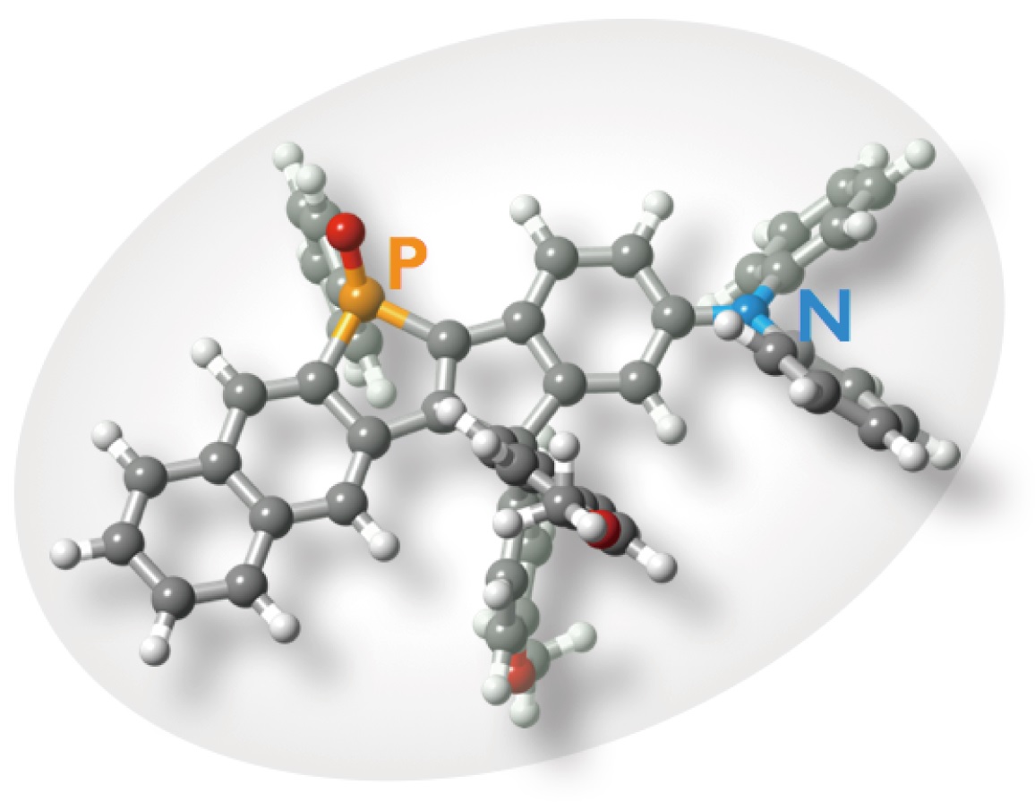
Molecular structure of C-Naphox.
Optical microscopy allows researchers to see and distinguish between objects that are about 200 nanometres (nm) apart. In comparison, a human hair is about 90,000 nm thick. Unfortunately, most objects of interest in biology, such as organelles in cells and proteins, are much smaller than 200 nm.
Biologists have been looking for ways to improve the resolution of microscopes, pioneering the field of super-resolution microscopy. Stimulated emission depletion (STED) microscopy is one such improvement: a source of light focuses on a point of interest while the surrounding zone is kept in the dark and toned down, so to speak, using a special laser to form a background without interferences. This technique is fluorescence-based, using special dyes to tag the cells or structures of interest.
STED microscopy is very effective, allowing researchers to detect objects that are only tens of nanometres apart. However, it does come with its own set of challenges: most importantly, that the special laser used to tone down the background is, counter-intuitively, very intense. Not many dyes can withstand this intensity without losing fluorescence so quickly that only a few images can be taken, which is much too fast for the needs of researchers.
Professor Shigehiro Yamaguchi and Professor Tetsuya Higashiyama from the Institute of Transformative Bio-Molecules at Nagoya University in Japan have developed a dye, called C-Naphox, that, thanks to a carbon-bonded structure, is very stable and does not dim even under the harsh conditions of STED microscopy. It is also non-toxic, so it can be used in live cells.
The researchers found that the dye remained stable after two hours of irradiation. When taking multiple images in succession—a key part of super-resolution microscopy as it allows researchers to follow live cells undergoing their natural processes over time—the team found that C-Naphox remained stable after five images. Even after taking 50 images, more than 80% of the C-Naphox signal remained. In comparison, one of the best options available commercially, a compound called Alexa 488, dimmed almost to invisibility after taking only five images. Once widely available, C-Naphox should enable prolonged recording of live cells using STED microscopy; a previously unattainable feat.
Further information
Professor Shigehiro Yamaguchi | E-mail: [email protected]
Institute of Transformative Bio-Molecules (ITbM)
Nagoya University
Professor Tetsuya Higashiyama | E-mail: [email protected]
Institute of Transformative Bio-Molecules (ITbM)
Nagoya University
Meet the Expert
Yamaguchi works on a variety of topics in the general fields of main group chemistry and physical organic chemistry. In particular, emphasis is placed on the development of new functional π-electron materials possessing unusual photophysical and electronic properties. On the basis of the new design concepts emanating from his lab, he is able to exploit the features of various main group elements as well as the newly developed synthetic methodologies, various types of functional materials have been developed; including the key molecule in commercial organic electroluminescent devises. He will be involved mainly in the development of innovative bio-imaging tools, collaborating with the groups of Higashiyama, Irle, Itami, and Crudden.
Higashiyama is one of the most recognized plant biologists focusing on the identification of key molecules of plant reproduction. In particular, his discovery of the long-sought (~140 years) pollen tube attractant molecule "LURE", which is essential for plant reproduction, has secured his position as a world-renowned biologist. The identification of LURE peptides provide us with a major breakthrough to study and control pollen tube guidance of flowering plants and to break reproductive barriers. Moreover, he has also made significant technical development of micro-genomics and live cell imaging. Currently, he serves as one of the youngest directors of prestigious ERATO funding program supported by the Japan Science and Technology Agency. He will be involved mainly in the development of molecules that can overcome species barriers as well as innovative bio-imaging tools, collaborating with the groups of Yamaguchi, Irle, Torii, Itami, Ooi, Crudden, and Bode.
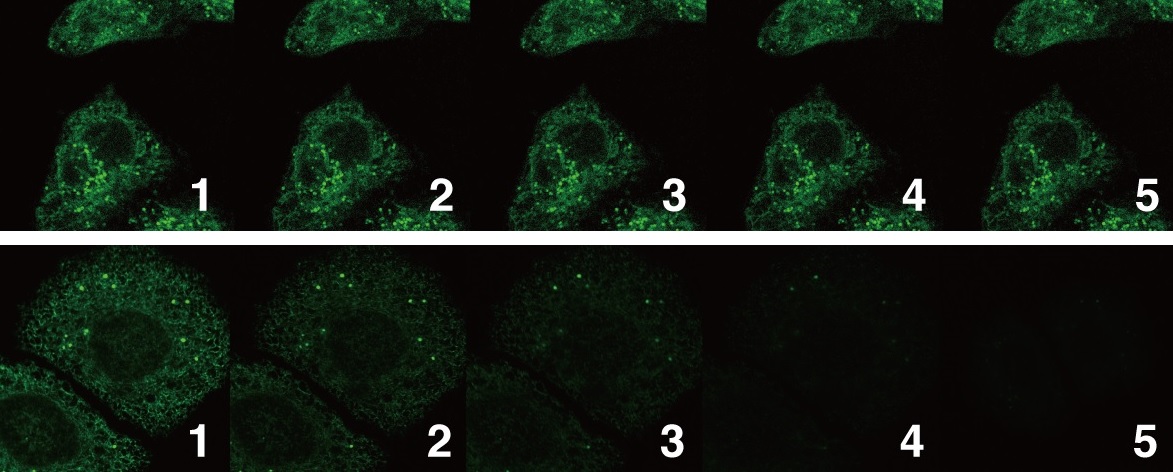
(Above) STED microscopy images of cells with C-Naphox remained stable. (Below) STED microscopy images with a commercially available compound.
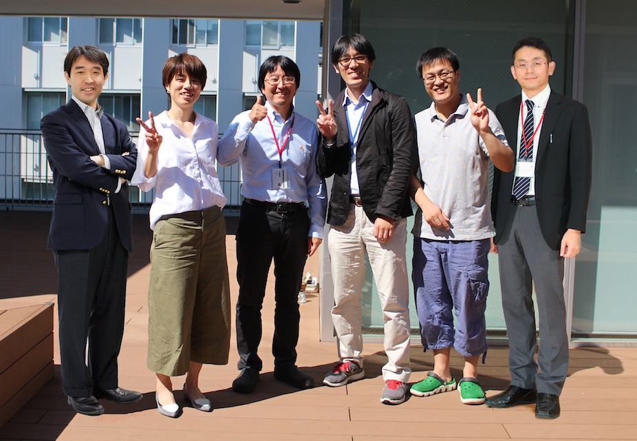
C-Naphox inventors from left: Tetsuya Higashiyama, Aiko Fukazawa, Yoshikatsu Sato, Masayasu Taki, Chenguang Wang, Shigehiro Yamaguchi.