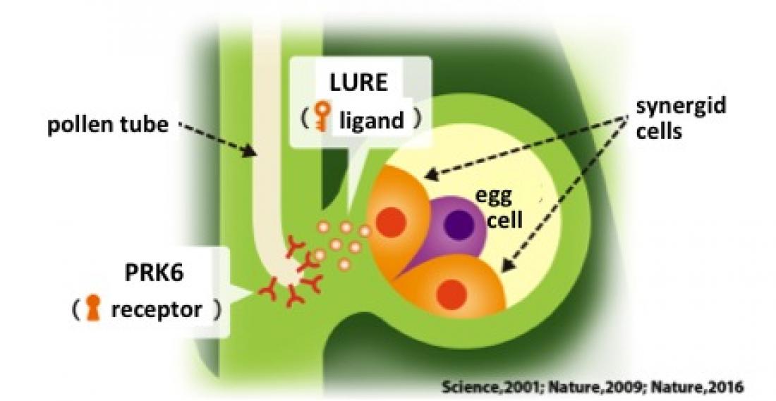
The LURE peptide, which is secreted by the synergid cells within the ovule acts as a key to bind to the lock, which is the PRK6 receptor found on the tip of the pollen tube. Figure taken and adapted from the webpage of "The birth of new plant species", a project supported by the Grant-in-Aid for Scientific Research on Innovative Areas (http://www.ige.tohoku.ac.jp/prg/plant/).
~ Solving the cocrystal structure of a pollen tube attractant and its receptor ~
Pollen tube guidance towards the ovule is an important step for fertilization in flowering plants. In order for this to happen, a pollen tube attractant peptide LURE guides the pollen tube precisely to the ovule. An international team of plant biologists at Nagoya University and Tsinghua University has succeeded in analyzing for the first time, the crystal structure of LURE bound to its receptor protein PRK6. Further elucidation of this key and lock mechanism may lead to applications in generating useful hybrid plant species.
Nagoya, Japan – Fertilization in flowering plants occurs by the delivery of sperm cells to the ovule by the precise growth of pollen tubes from pollen. Pollen tube guidance plays a crucial role in controlling the growth of pollen tubes and a pollen tube attractant peptide LURE is secreted from the synergid cells next to the egg cell within the ovule to lead to successful fertilization. LURE is specific to each plant species and is therefore responsible for the fertilization between the same species.
LURE1 has already been identified in a model plant Arabidopsis thaliana, and there have been reports on the presence of receptors on the pollen tube responsible for detecting LURE1. The key and lock model illustrates the relationship between the LURE peptide (ligand) and its receptor. To which lock (receptor) the key (LURE) binds to and how it does so has been a mystery up to now.
In order to identify the exact receptor on the pollen tube for LURE, Tetsuya Higashiyama, a professor at Nagoya University and his collaborators at Tsinghua University who have expertise in structural biology of plant ligands and receptors, performed analyses of the complexes by X-ray crystallography. The team examined the protein that binds to LURE by making LURE of Arabidopsis thaliana and its protein receptor by cultures of insect cells. As a result, they were able to determine that LURE specifically binds to a protein receptor called PRK6 (pollen receptor-like kinase 6) on the pollen tube. The results of this study are reported in Nature Communications.
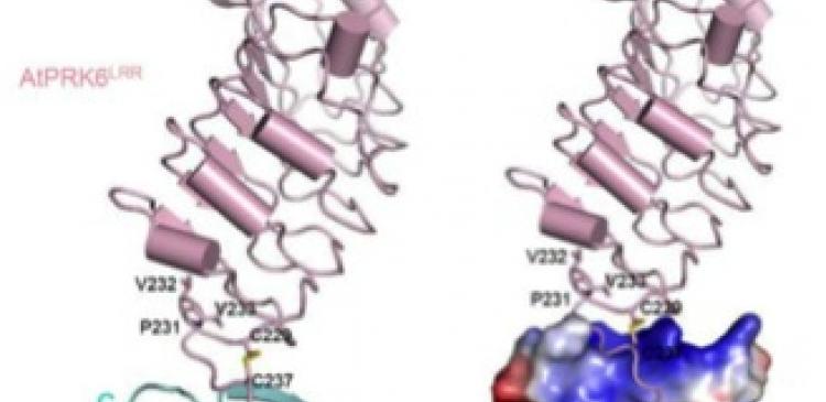
The research team succeeded in obtaining and analyzing the crystal structure of LURE bound to the PRK6 receptor. As a result of their analyses, they found that LURE is bound by being inserted between the leucine-rich repeat region and the transmembrane region of the PRK6 receptor.
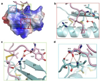
LURE is bound to a region near the membrane of PRK6. This region is called the loop region and the team identified that the electrostatic interaction between the positive charge of LURE and the negative charge on the loop region of PRK6 was significant for the binding. In addition, they were also able to show that a disulfide bridge is formed in the PRK6 loop region upon the binding of LURE. This bridge between cysteine residues contributes to the stabilization of the loop region that plays a significant role for the binding to LURE.
Previous reports have shown that binding between peptides and receptors in plants mainly occurs at the leucine-rich repeat region. Secondly, when a ligand molecule binds to a receptor on the outer region of the cell, a complex is usually formed to communicate signals within the cell. On the other hand, the binding between LURE and PRK6 had occurred at a different position to what has been reported in plants so far, and no complexes were made upon binding. This unique binding scheme between LURE and PRK6 appears to reflect the precise control mechanism of the direction of pollen tube growth.
Upon having a closer look into the binding between LURE and loop region of PRK6, they were able to identify the relevant amino acids required for the binding. The team tested the degree of binding and pollen tube guidance by switching around the amino acids, and found that both arginine (R83) located in LURE and aspartic acid (D234) in PRK6 were important.
“Our lab has worked intensively to identify the pollen tube attractant peptide LURE and its receptor protein PRK6, and this time, we were able to determine the crystal structure of the LURE bound complex to prove that PRK6 is the actual receptor of LURE,” says Higashiyama.
“Through its interaction with the PRK6 receptor, we found that LURE is able to change the direction of pollen tube growth towards the ovule, but we are still unsure of how LURE is able to control the growth direction with such high precision,” explains Higashiyama. “We have demonstrated that PRK6 tends to concentrate towards LURE, so it may be that upon the binding of LURE in the loop region of PRK6, PRK6 changes its behavior on the pollen tube tip. By carrying out real time observations of the molecular activities of LURE and PRK6 on the surface of pollen tubes, we hope to understand the exact mechanism of pollen tube guidance.”
The LURE peptide and the PRK6 receptor act as a key and lock, which is specific for each plant species. Higashiyama and his group envisage that this work will help to reveal why pollen tube guidance is successful between the same species and is difficult between different species.
“We hope to be able to design specific ‘key and lock’ systems so that pollen tube guidance becomes efficient between different plant species, and can lead to successful hybridization between different species,” says Higashiyama. “Plants such as bread wheat, rape seed, and cotton are all important species which have arisen as a result of cross-breeding. We believe that our research will be significant towards the generation of specifically designed cross-breeding plant species.”
This article “Structural basis for receptor recognition of pollen tube attraction peptides” by Xiaoxiao Zhang, Weijia Liu, Takuya T. Nagae, Hidenori Takeuchi, Heqiao Zhang, Zhifu Han, Tetsuya Higashiyama & Jijie Chai is published online in Nature Communications. DOI: 10.1038/s41467-017-01323-8 (http://dx.doi.org/10.1038/s41467-017-01323-8)
Author Contact
Professor Tetsuya Higashiyama
Institute of Transformative Bio-Molecules (WPI-ITbM), Nagoya University
Furo-cho, Chikusa-ku, Nagoya 464-8601, Japan
TEL: +81-52-747-6404 FAX: +81-52-747-6405
E-mail: [email protected]
Media Contact
Dr. Ayako Miyazaki
Institute of Transformative Bio-Molecules (WPI-ITbM), Nagoya University
Furo-cho, Chikusa-ku, Nagoya 464-8601, Japan
TEL: +81-52-789-4999 FAX: +81-52-789-3053
E-mail: [email protected]
About WPI-ITbM (http://www.itbm.nagoya-u.ac.jp/)
The Institute of Transformative Bio-Molecules (ITbM) at Nagoya University in Japan is focuses on advancing the integration of synthetic chemistry, animal/plant biology and theoretical science, all of which are traditionally strong fields in the university. ITbM is one of the research centers of the Japanese MEXT (Ministry of Education, Culture, Sports, Science and Technology) program, the World Premier International Research Center Initiative (WPI). The aim of ITbM is to develop transformative bio-molecules, innovative functional molecules capable of bringing about fundamental change to biological science and technology. Research at ITbM is carried out in a "Mix Lab" style, where international young researchers from various fields work together side-by-side in the same lab, enabling interdisciplinary interaction. Through these endeavors, ITbM will create "transformative bio-molecules" that will dramatically change the way of research in chemistry, biology and other related fields to solve urgent problems, such as environmental issues, food production and medical technology that have a significant impact on the society.


