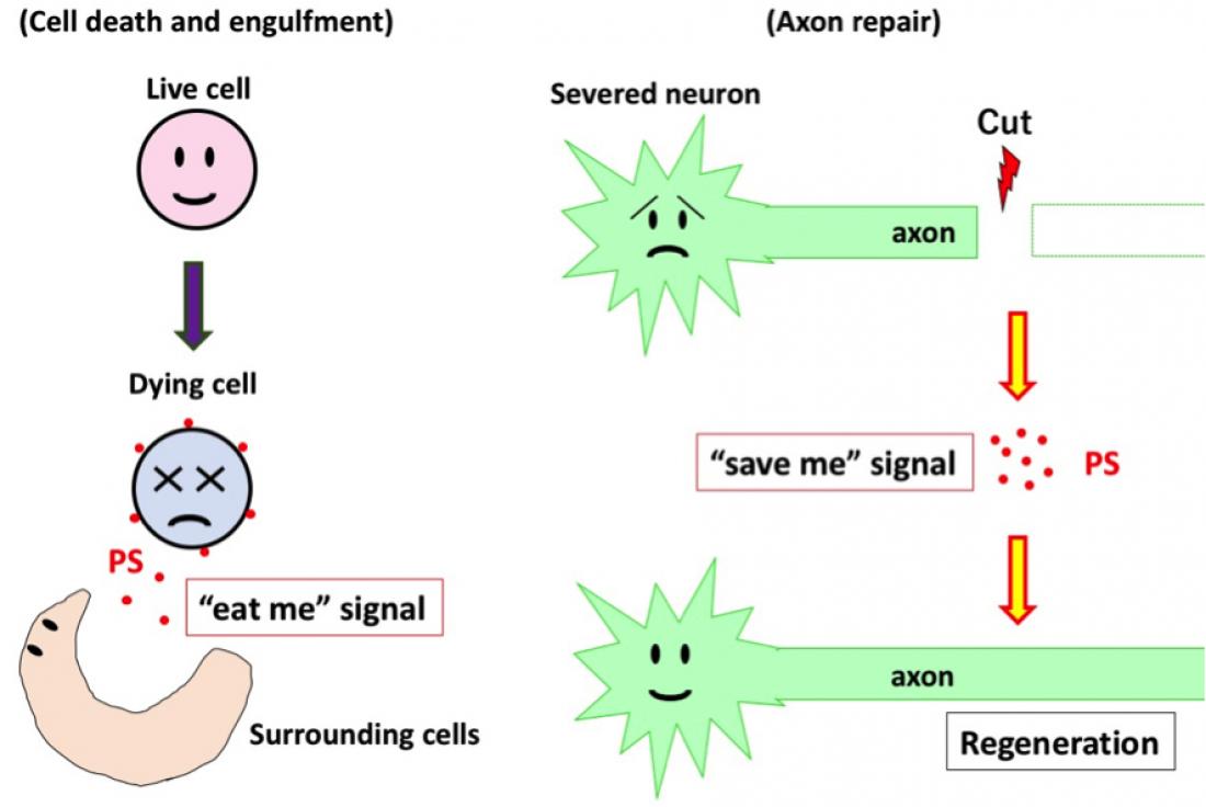
The dying cells secrete phosphatidylserine (PS), a kind of lipid acting as an “eat me” signal, into the extracellular space. PS is then recognized by the engulfing cells that eat the dying cells. The severed neurons, however, induce only the PS secretion without dying. The secreted PS then acts as a “save me” signal that accelerates axon regeneration of the severed neurons.
Nagoya, Japan - The branches of nerve cells called axons are particularly susceptible to damage due to the long distances they extend to communicate with each other. In humans, such damage in peripheral regions of the body can be relatively well repaired, but this repair is less effective in the brain and the spinal cord, which helps to explain why conditions such as brain and spinal cord injuries are so debilitating. In a new paper published in the journal Nature Communications, researchers at Nagoya University have made a major advancement in characterizing how axons regenerate by studying the roundworm Caenorhabditis elegans, a species that is widely used in biological research and has a very well-characterized nervous system. Specifically, they have shown that axon repair occurs using largely the same set of molecules that mediate the recognition and engulfment of apoptotic (dying) cells by the surrounding cells. The result suggests that this system has been co-opted for an additional purpose over the course of evolution. The team used a laser to cut roundworm axons and then analyzed the subsequent series of molecular reactions that occurred. They found that this damage resulted in the movement of a lipid called phosphatidylserine (PS) from the inside of cells to their outside, which was mediated by a protein called an ABC transporter. This externalized PS was then recognized by another molecule, triggering a series of reactions that eventually led to repair of the axon. Interestingly, PS is better known as an "eat me" signal that helps the phagocytosis of a dying cell by its neighbors. "We were able to dissect the complex range of molecules involved in axon repair by using fluorescent labels in and around the severed axon and knocking down the individual components suspected of being involved," says corresponding author Kunihiro Matsumoto. "Although many of these molecules are also active in promoting phagocytosis of apoptotic cells, in axon repair that creates a 'save me' signal rather than an 'eat me' one, which enables the axons to regenerate." The team explains that for the repair of damaged nerves, the PS labeling appears only at the severed sites and exists for only a short time (~1 hr), which is in contrast to the labeling in eliminating dying cells that remains for a long time until the cells are eliminated. The researchers now guess that this difference in signal timing may be one way for the cells to distinguish the meaning of the PS signal - 'eat me' vs. 'save me.' According to Naoki Hisamoto, "Now that we know how this system works in the relatively simple roundworm, we should eventually be able to extrapolate the findings to humans. This could provide us with a range of targets for pharmaceutical interventions to treat conditions like brain and spinal cord injuries, in which the human body is not able to repair damaged nerves."


