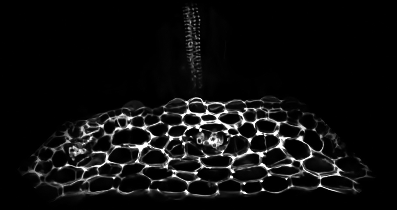
Reconstructed image of a honeycomb structure of lignin in sepals, the leaf-like part of the plant that protects the flower bud.
Plants shed leaves, flowers, fruits or diseased parts in various developmental stages, but the underlying mechanisms of how this happens was unclear. Now, research has identified two cell types that execute the precise separation of flowers in the model plant Arabidopsis. The findings provide a basic mechanism for the plant cell separation process, and have implications for crop improvement strategies.
Unlike animals, plants have rigid cell walls that strongly bond to each other. While this provides a solid outer structure, it is also a physical constraint for plants undergoing physiological changes, such as cell expansion, wound healing and responses to their surrounding environment. For example, wound healing in animals involves cell migration, but in plants, cell walls need to be rebuilt.
Past studies identified some of the key players involved in cell wall loosening and degradation to help initiate floral separation in Arabidopsis. However, how the actual shedding occurs with great precision was unknown.
In a Cell study, plant biologist June Kwak of South Korea’s Daegu Gyeongbuk Institute of Science and Technology (DGIST) and his colleagues identified two different cell types in the ‘abscission zone’ that conduct the final stage of cell wall separation in a finely tuned manner: residuum cells (RECs) in the main plant body and secession cells (SECs) in the separating part.
They discovered that SECs form a honeycomb-shaped structure of lignin, a phenolic polymer in cell walls that makes plants rigid. Lignin helps limit the work of enzymes that degrade the cell wall to the shedding area, and simultaneously clutches the separating cells like a molecular brace until the moment of shedding, ultimately falling off with the separated organ.
The shedding process triggered by the lignin leads to RECs transforming into epidermal cells, forming a protective coating on the main body where the flower has just been shed. This prevents bacterial infections on the new surface.
The transformation from non-epidermal cells into epidermal cells was previously thought to occur only in embryogenesis at the beginning of a plant’s life cycle. Organ separation might provide a new window for studying epidermal cell specification, which had been hindered by their unstable state in early developmental stages. In particular, this could help researchers examine how RECs transform into epidermal cells, what affects such transformations, and how neighbouring cells communicate with each other.
The researchers hope that the information obtained in this study on floral separation may help in the understand- ing of other cell separation events in different developmental phases of plants. Furthermore, the finding could help improve crop productivity if researchers are able to control the timing of shedding flowers, fruits, and leaves. Future research should also look at the difference between sealing accidental wounds versus organ shedding.
For more information, contact:
Professor June M. Kwak
Department of New Biology
Daegu Gyeongbuk Institute of Science and Technology
E-mail: [email protected]