


Researchers from National Taiwan University and Taipei Veterans General Hospital developed an AI system that automatically measures spinal curvature from X-rays. The system remained accurate when tested on images from a different institution, showing potential for more consistent scoliosis assessment across hospitals.
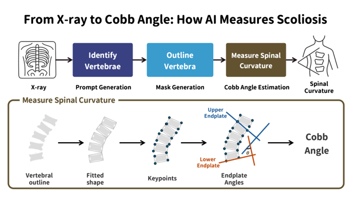
The AI system automatically identifies individual vertebrae, outlines their shapes, and calculates spinal curvature from X-ray images.
National Taiwan University
Scoliosis is a condition characterized by an abnormal sideways curvature of the spine and most commonly develops during adolescence. Doctors typically assess its severity by measuring spinal curvature on X-ray images. This measurement plays an important role in diagnosis, follow-up evaluation, and treatment planning.
However, it is still largely performed manually, making the process time-consuming and subject to differences between clinicians and between repeated measurements by the same clinician. Part of this variability can arise from how the vertebrae defining the spinal curve are selected.
Researchers from National Taiwan University and Taipei Veterans General Hospital have developed an artificial intelligence system designed to make scoliosis measurement more automatic and consistent. The system analyzes a spinal X-ray in several steps. It first identifies each vertebra and examines its shape and tilt to automatically measure spinal curvature. The study is published in IEEE Transactions on Medical Imaging.
The researchers evaluated the system using spinal X-rays from Taipei Veterans General Hospital. Compared with measurements made by experienced specialists, the AI showed strong agreement. In cases where the two human observers differed by no more than five degrees, the average differences between AI and specialist measurements ranged from approximately 2.3 to 3.4 degrees across the evaluated spinal curves. Overall, the agreement between the AI and doctors was comparable to the agreement between two human observers.
An important part of the study was testing whether the system could also work on X-rays from a different source. Medical images can vary across hospitals because of differences in imaging equipment, acquisition procedures, and overall image appearance. The researchers therefore evaluated the system on an external dataset collected under different institutional and imaging conditions. Although the AI was not retrained on these external images, it continued to perform well, suggesting its potential for broader use across different hospitals and clinical settings.
Taken together, the findings suggest the system could serve as a decision-support tool to help doctors obtain more reproducible scoliosis measurements while reducing the time and variability associated with manual assessment.
“Our goal is not to replace clinical judgment, but to provide doctors with a reliable tool that can make scoliosis measurement more consistent and reduce the variability that can occur during manual assessment,” says co-corresponding author I-Yun Lisa Hsieh, associate professor of civil engineering at National Taiwan University.
Prof. I-Yun Lisa Hsieh’s email address: [email protected]
Advertisement