
To the left, Professor Jung-Ah Cho, who helped conceptualize the study. In the middle and to the right, lead authors Ms Yoojin Ahn and Mr Tae Seong Lyu, respectively.
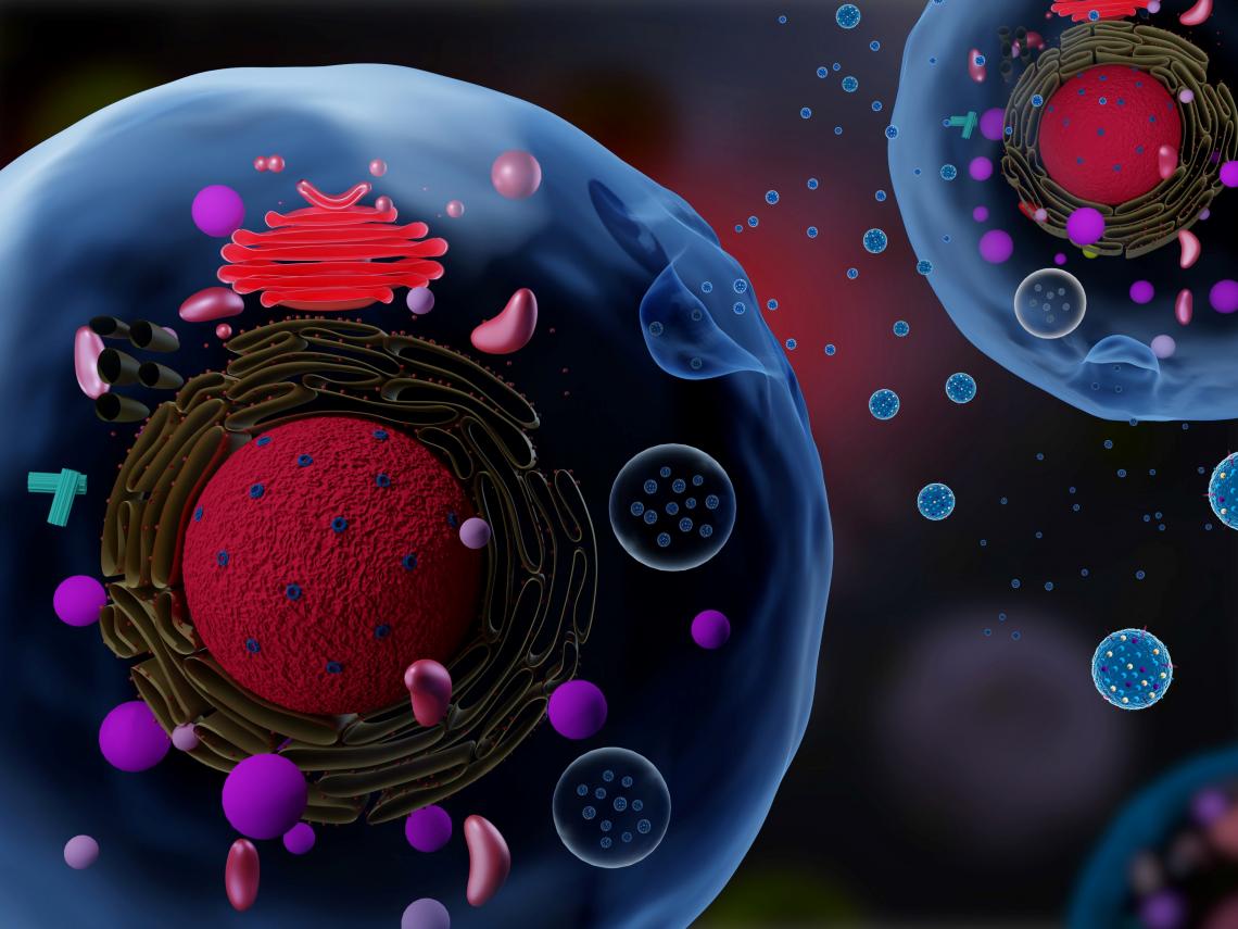
Exosomes are small vesicles released by cells that are thought to be important for cell communication and regulation. They are likely to play a role in tumor progression and cancer as well
Exosomes are small vesicles released by cells, which contain proteins and genetic materials. They are thought to be involved in various life activities, but current techniques to observe them are expensive and time-consuming. In a recent study, a team of undergraduate students from DGIST, Korea, proved the convenience and effectiveness of laser-based technique in identifying exosomes using exosomes from a less studied cancer type, paving the way for future exosome research.
Despite our great progress in understanding various cellular mechanisms over the last decades, many of them remain unclear. Such is the case for exosomes, small vesicles released by cells that contain genetic materials called “RNA” and various proteins. The roles of exosomes are believed to be very varied and important, both for normal bodily functions and also in the spreading of diseases like cancer. However, exosomes are so small that studying them is challenging and calls for costly and time-consuming techniques, such as electron microscopy (EM).
To tackle this issue, a team of undergraduate students from Daegu Gyeongbuk Institute of Science and Technology (DGIST), Korea, explored a different and promising method for analyzing exosomes. In their study, which was published in PLOS One, they focused on dynamic light scattering (DLS), a laser-based technique that can be used to easily determine statistical parameters about the sizes of a large number of vesicles. What was admirable, as per Prof Jung-Ah Cho (corresponding author of the study), was that “the undergraduate students independently conducted the whole study under DGIST’s Undergraduate Group Research Program with no external help.”
First, they compared the exosomes of two types of cancer cells: a well-studied breast cancer cell line and a mouse fibrosarcoma cell line. The exosomes secreted by cancer cells of the latter type had rarely been studied before. Thus, to bring more information to the table, the team also used EM and other analytical techniques (such as Western blot) in addition to DLS.
Aside from identifying proteins within the exosomes, the results helped clarify the advantages and disadvantages of EM and DLS. Overall, when used properly, DLS seems to be a reliable and convenient tool for studying exosomes, as Prof Cho explains: “DLS is an efficient technique that is comparatively inexpensive and simple; it can quickly and sensitively measure the size and purity of exosomes without technical difficulties. In contrast, EM is expensive and not suitable for daily or routine measurements.”
Having a more accessible technique to study exosomes should help scientists all over the world get involved in exosome research. When asked about the exact implications of using DLS for this purpose, Prof Cho replied, “The many advantages of DLS for analyzing exosome extracts could speed up research on disease diagnosis, prognosis monitoring, and drug delivery systems using exosomes.” From a more academic point of view, the results of this study provide some of the basic data needed to understand the biological activities of exosomes derived from fibrosarcoma.
This study represents a significant step towards a better understanding of exosomes and their roles, both in sickness and in health. With any luck, DLS will play its part to solve these mysteries.
Reference
|
Authors: |
Tae Seong Lyu1, Yoojin Ahn1, Young-Jun Im1, Seong-Soo Kim1, Ki-Heon Lee1, Jinyoung Kim1, Yujin Choi1, Dongwoo Lee1, EunSeok Kang1, Gayeon Jin1, Jiwon Hwang1, Sang-im Lee1,2, Jung-Ah Cho1 |
|
Title of original paper: |
The characterization of exosomes from fibrosarcoma cell and the useful usage of Dynamic Light Scattering (DLS) for their evaluation |
|
Journal: |
PLOS One |
|
DOI: |
10.1371/journal.pone.0231994 |
|
Affiliations: |
1College of Transdisciplinary Studies, School of Undergraduate Studies, DGIST 2Department of New Biology, Graduate School, DGIST |
*Corresponding author’s email: [email protected]
About Daegu Gyeongbuk Institute of Science and Technology (DGIST)
Daegu Gyeongbuk Institute of Science and Technology (DGIST) is a well-known and respected research institute located in Daegu, Republic of Korea. Established in 2004 by the Korean Government, the main aim of DGIST is to promote national science and technology, as well as to boost the local economy.
With a vision of “Changing the world through convergence", DGIST has undertaken a wide range of research in various fields of science and technology. DGIST has embraced a multidisciplinary approach to research and undertaken intensive studies in some of today's most vital fields. DGIST also has state-of-the-art-infrastructure to enable cutting-edge research in materials science, robotics, cognitive sciences, and communication engineering.
Website: https://www.dgist.ac.kr/en/html/sub01/010204.html
About the authors
All the undergraduate students who were part of the Undergraduate Group Research Program cooperated well and were spontaneously involved. They also divided and performed their roles properly. Tae seong Lyu and Yoojin Ahn led this DLS research, from planning and performing experiments and analyzing data to writing the manuscript draft. The other team members, Young-Jun Im, Seong-Soo Kim, and Ki-Heon Lee, studied the molecular characteristics of exosomes through molecular and biochemical experiments. In addition, other students volunteered to participate in this work during the revision; they accomplished their designated roles through cooperation. Prof Sang-im Lee contributed to completing the manuscript.