Raman image of rapidly frozen HeLa cells with high signal-to-noise ratio and large field-of-view. The image acquisition time was 10 hours. The distribution of Raman signals from cytochromes (750 cm⁻¹), lipids (2850 cm⁻¹), proteins (2920 cm⁻¹), are indicated in green, red, and blue, respectively.
Researchers from Osaka University use cryogenic freezing to achieve high-resolution Raman microscopy images of biological samples
Osaka, Japan – Understanding the behavior of the molecules and cells that make up our bodies is critical for the advancement of medicine. This has led to a continual push for clear images of what is happing beyond what the eye can see. In a study recently published in Science Advances, researchers from Osaka University have reported a method that gives high-resolution Raman microscopy images.
Raman microscopy is a useful technique for imaging biological samples because it can provide chemical information about specific molecules—such as proteins—that take part in the body’s processes. However, the Raman light that comes from biological samples is very weak, so the signal can often get swamped by the background noise, leading to poor images.
The researchers have developed a microscope that can maintain the temperature of previously frozen samples during the acquisition. This has allowed them to produce images that are up to eight times brighter than those previously achieved with Raman microscopy.
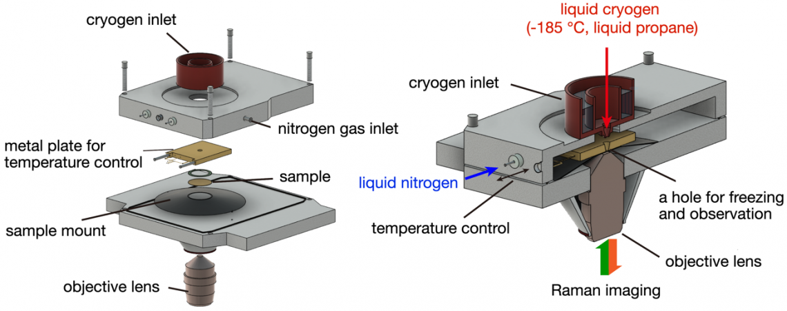
Schematic of the sample area in the developed cryo-Raman microscope. A cover glass with cultured cells is placed on the sample mount, and a metal plate is brought into contact (left). The cryogen enters through the inlet and a hole in the metal plate, coming into direct contact with the sample for rapid freezing. The sample temperature is controlled by the metal plate with liquid nitrogen circulation and a heater inside (right).
“One of the main reasons for blurry images is the motion of the things you’re trying to look at,” explains lead author of the study, Kenta Mizushima. “By imaging frozen samples that were unable to move, we could use longer exposure times without damaging the samples. This led to high signals compared with the background, high resolution, and larger fields of view.” The technique uses no stains and doesn’t require any chemicals to fix the cells in position, so can provide a highly representative view of processes and cell behavior.
The team was also able to confirm that the freezing process conserved the physicochemical states of different proteins. This gives the cryofixing approach a distinct advantage of achieving what the chemical fixing methods cannot.
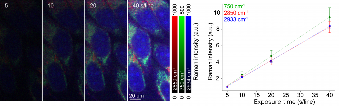
Raman images of rapidly frozen HeLa cells captured with different exposure times (left) and the relationship between the Raman signal intensity, attributed to different molecular vibrations, and exposure time (right). The observation temperature was -40°C.
“Raman microscopy adds a complementary option to the imaging toolbox,” says senior author Katsumasa Fujita. “The fact that it not only provides cell images, but also information about the distribution and particular chemical states of molecules, is very useful when we are continually striving to achieve the most detailed possible understanding.”
The new technique can be combined with other microscopy techniques for detailed analysis of biological samples and is expected to contribute to a wide range of areas in the biological sciences including medicine and pharmaceutics.
###
The article, “Raman microscopy of cryofixed biological specimens for high-resolution and high-sensitivity chemical imaging,” was published in Science Advances at DOI: https://www.science.org/doi/10.1126/sciadv.adn0110
About Osaka University
Osaka University was founded in 1931 as one of the seven imperial universities of Japan and is now one of Japan's leading comprehensive universities with a broad disciplinary spectrum. This strength is coupled with a singular drive for innovation that extends throughout the scientific process, from fundamental research to the creation of applied technology with positive economic impacts. Its commitment to innovation has been recognized in Japan and around the world. Now, Osaka University is leveraging its role as a Designated National University Corporation selected by the Ministry of Education, Culture, Sports, Science and Technology to contribute to innovation for human welfare, sustainable development of society, and social transformation.
Website: https://resou.osaka-u.ac.jp/en