The research team is led by Dr Brian Kot Chin-wing, Visiting Assistant Professor of the Department of Veterinary Clinical Sciences and Research Associate of the State Key Laboratory of Marine Pollution (SKLMP) at CityU. Their findings have been published in the scientific journal Frontiers in Marine Science, titled “Virtopsy as a Revolutionary Tool for Cetacean Stranding Programs: Implementation and Management”.
Non-invasive virtopsy: more accurate and time-saving
Virtopsy, also known as postmortem imaging, is the examination of dead bodies with modern imaging modalities before conventional necropsy (similar to “body-opening” autopsy in humans). “Virtopsy appears to be more accurate, time-saving, and non-invasive when compared to conventional necropsy, with a lesser risk of disease contraction for veterinarians and other human rescuers,” said Dr Kot.
“Most of the cetacean carcasses stranded in Hong Kong were badly decomposed. Therefore, full necropsy could not be conducted. As a result, the cause of death of 90% of cases remained undetermined and important biological health concerns, for example, amount of parasites inside the body and related pathogenic effects on the hosts’ fitness, would be missed,” he explained.
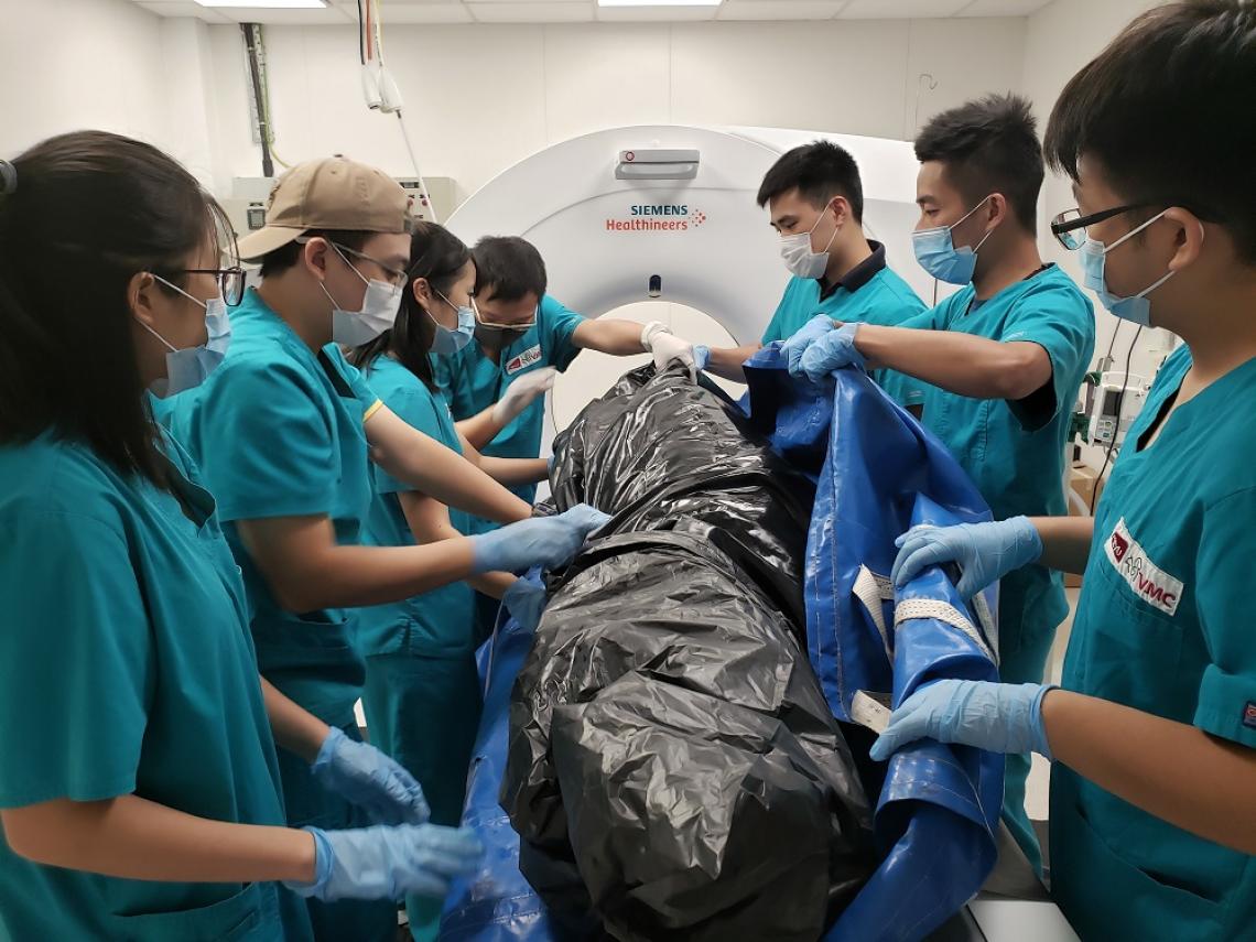
Dr Kot and his team are preparing the dead body of a Chinese white dolphin for virtopsy. (Photo source: Dr Brian Kot)
Under such circumstances, virtopsy is a good alternative that may give invaluable initial or additional information by documenting the dead bodies’ external and internal structures using high throughput imaging techniques. This can facilitate the investigation of the cause and manner of death, as well as other important biological health concerns. More importantly, there is no need to open up the dead bodies, which is complex and time-consuming.
Pioneer Cetacean Virtopsy Stranding Response Programme management scheme
The CityU team led by Dr Kot have been working hand in hand with the Hong Kong Agriculture, Fisheries and Conservation Department (AFCD), Ocean Park Corporation (OPC), Ocean Park Conservation Foundation Hong Kong (OPCFHK) in a pioneer Cetacean Virtopsy Stranding Response Programme management scheme in Hong Kong waters for many years. The team initiated virtopsy scanning on the stranded cetaceans in 2014 and became the first team worldwide to conduct virtopsy routinely.
The scheme works like this: After receiving a stranding report, AFCD would refer the case to OPCFHK. Then OPCFHK would arrange a pick-up and record the information on-site, including the stranding time and location, size, decomposition condition, and presence of wounds. Due to scanning modalities limitation, virtopsy could not be done on cetaceans heavier than 250 kg and those bodies’ diameter exceeded 90 cm. These bodies would not be transported. Instead, on-site necropsy would be performed and samples would be collected. Bodies that could be transported would be sent for virtopsy by Dr Kot and his team in the Aquatic Animal Virtopsy Lab. After that, necropsy would be performed by OPC and OPCFHK.
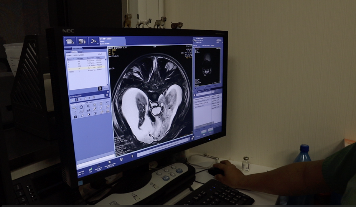
Dr Kot’s team examines the virtopsy images. (Photo source: Dr Brian Kot)
Dr Kot and his team have reviewed the scheme and identified some pitfalls. They proposed some practical suggestions to address them in facilitating the entire workflow. For example, they suggested that a one-stop examination centre should be established, integrating advanced technologies and standard examination methods, including virtopsy and conventional necropsy. This could reduce the time and resource-demanding transportation.
“Our study is a valuable reference for other stranding response programmes worldwide that are interested in integrating virtopsy or other modern diagnostic modalities into their routine workflow. We hope our research could help not only the aquatic animals but also human beings to realise a 'One Ocean-One Health' ideal,” conclude Dr Kot.
A comparison between PMCT and PMMRI
Dr Kot and his team have already examined 230 local stranded cetaceans by virtopsy using postmortem computed tomography (PMCT) and postmortem magnetic resonance imaging (PMMRI), providing vital or additional information other than conventional necropsy.
By further comparing the two techniques on the investigation of the brains of 18 carcasses, Dr Kot and his team found that PMCT allowed excellent identification of bone lesions, foreign bodies, pathological gas formation, and organ trauma with higher precision over the conventional autopsy. On the other hand, PMMRI performed better in demonstrating soft tissue injury, neurological and nonneurological organ trauma and non-traumatic pathology.
It is common for stranded carcasses to show rapid cerebral autolysis and putrefaction, due to their high water content. But these decomposed brains could still provide useful information on biological health concerns of the stranded cetaceans. “Our study demonstrated that even for decomposed brains, PMMRI could offer a more distinct soft tissue contrast and allow detailed morphological assessment when compared to PMCT. We thereby recommended that brain PMMRI should be incorporated in the postmortem investigation of decomposed cetacean carcasses,” said Dr Kot.
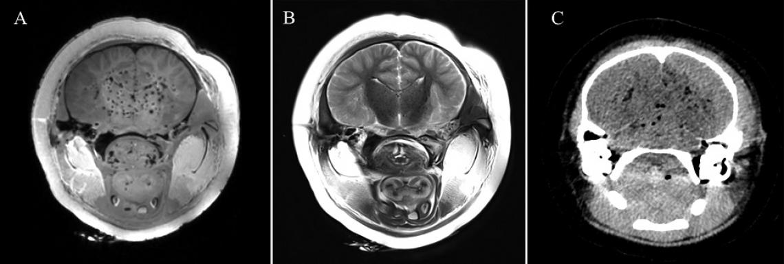
Despite gas accumulation in the decomposed cetacean brain, various neuroanatomical structures are still well demonstrated in both PMMRI images (Figure A & B), compared to the PMCT image (Figure C) of the same section. (Photo source: DOI number: 10.3389/fmars.2020.544037)
Also, the team suggested using PMMRI in complementarily with PMCT in detecting soft tissue lesions and parenchymal pathologies in the brain, as PMMRI alone is more prone to artefacts from gas and foreign bodies.
The team hoped that these findings could help in providing better insights into the health condition of cetacean brains, and revealing more secrets of the intelligence that many cetaceans possess. Moreover, Dr Kot pointed out that documentation of brain morphology could also help to investigate emerging threats to stranded cetaceans such as anthropogenic chemicals and biotoxins from algal blooms.
Such findings have also been published in the scientific journal Frontiers in Marine Science, titled “Postmortem Neuroimaging of Cetacean Brains Using Computed Tomography and Magnetic Resonance Imaging”.
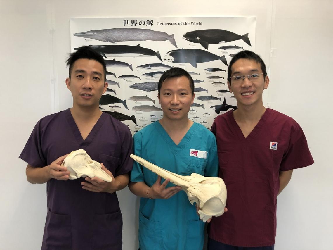
Dr Kot (middle) and his team members: Dr Tabris Chung (left) and Mr Henry Tsui (right). (Photo source: Dr Brian Kot)
Dr Kot is the corresponding and first author of both papers. For the first paper, another co-first author is Mr Henry Tsui Chun-lok, Research Assistant from SKLMP at CityU. He is also a co-author in the second paper. Dr Tabris Chung Yik-to, also a Research Assistant from SKLMP, participated in both researches. The other collaborating researcher is from Tung Wah College.
Both studies received funding support from the Hong Kong Research Grants Council and the Marine Ecology & Fisheries Enhancement Funds Trustee Limited.
DOI number: 10.3389/fmars.2020.542015 and 10.3389/fmars.2020.544037