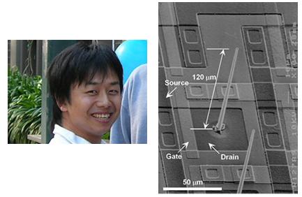
Dr Kawano and Fig.1: The 'Toyohashi Probe': An integrated VLS-silicon microprobe.
In nanotechnology, the so-called vapor-liquid-solid (VLS) method is widely used for synthesizing a variety of one-dimensional wire-structures including carbon nanotubes, and metallic and semiconducting nanowires for fabricating nanodevices.
Although the VLS method enables batch fabrication of out-of-plane vertically aligned micro and nanowires, a potentially powerful device application for measuring multi-site electrical neural signals has yet to be realized due to: (1) the unavailability of an device process for integrating the three-dimensional wire arrays with active devices; (2) inadequacies in the electrical properties of the tiny wires that are used as probes for recording electrical neural signals; and (3) the lack of an appropriate device packaging process that is compatible with saline.
Here, Takeshi Kawano, Makoto Ishida and colleagues at the Toyohashi University of Technology, Chukyo University, and RIKEN successfully demonstrate the neural recording capability of micrometer sized VLS-silicon wires—'Toyohashi Probe' using the retina of a fish (Fig.1 and Animation).
The researchers produced vertically aligned microprobe arrays on a silicon microelectronics substrate by a selective VLS growth of silicon followed by micro-fabrication processing and device packaging. For actual measurements the group placed the retina onto the Toyohashi Probes. These devices successfully detected neural responses representative of local field potentials of the retina (Fig.2).
Toyohashi Probes made by VLS growth show potential as powerful devices for a range of neural recordings because of the advantageous small sizes of the probes and their compatibility LSI electronics.
For references and the animation video, please click on the Toyohashi Tech eNewsletter below
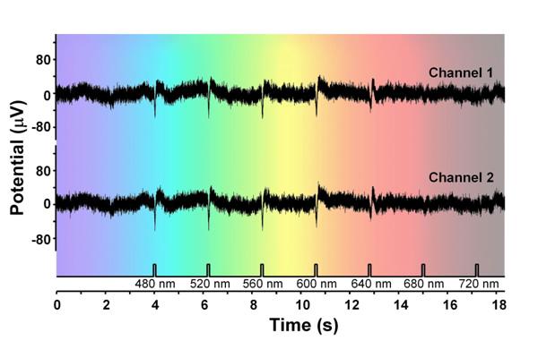
Fig. 2: Light-evoked neural signals of the retina (electro-retinogram (ERG)) measured via two probes.