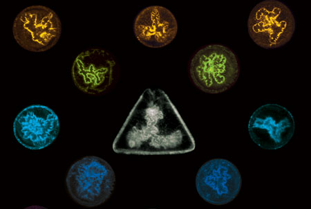
Fig 1. A composite image of epithelial tubes formed between the contacting lateral surfaces of two liver cells on artificial membranes in circular and triangular microwells. Researchers found that by coating the microwells with fibronectin in different patterns, the shape of the epithelial tubes that grow in the microwells are altered.
Singapore, 23 February 2016 – A team of scientists from Singapore and France has revealed the underlying mechanism for the formation and growth of a fundamental type of tissue – epithelial tubes. Defects in the architecture of epithelial tubes lead to diseases such as cholestasis, atherosclerosis and polycystic kidney disease. The research findings contribute towards a deeper understanding of the principles that underlie epithelial tube formation, and offer opportunities for developing better therapies for such diseases.
The study suggests that the shape and size of some types of epithelial tubes are governed by the mechanical forces that arise from the interaction of cells with the supportive extracellular matrix (ECM) that surrounds them. The work and its findings were published in the scientific journal Nature Cell Biology on 15 February 2016.
Extracellular matrix organization guides lumen morphology
All the major organs in the human body, such as the blood vessels, lungs, kidneys, liver, pancreas and the intestine, are formed of an extensive network of tubes. These tubes function as biological pipelines that transport and deliver life-sustaining liquids, gases or macromolecules from one site in the body to another. Depending on the organ in which they are formed or the specific function that they perform, the tubes vary greatly in size and shape, and defects in their tubular architecture have been linked to a number of diseases, such as atherosclerosis and polycystic kidney disease.
The tubes enclose hollow spaces called lumens and are primarily composed of a single or multiple layers of epithelial cells. As an important prerequisite for tube formation, epithelial cells become asymmetric or ‘polar’, acquiring structurally and functionally distinct ends or surfaces. Following this, cells undergo shape changes and organize around a central lumen, with their apical (top) surfaces facing the lumen, the basal (bottom) surfaces interacting with the underlying tissue and the lateral (side) surfaces in close contact with the neighboring cells. The vast majority of studies on tube formation have focused on understanding the molecular mechanisms leading to cell polarization and subsequent cellular mechanisms that drive the formation of lumens. However, factors that regulate the shape, size and the directional elongation of lumens into tubes remain unclear.
A recent collaborative study between Associate Professor Virgile Viasnoff, Principal Investigator at the Mechanobiology Institute (MBI) at the National University of Singapore and CNRS (France) and Professor Hanry Yu, Principal Investigator at MBI and Group Leader at the Institute of Bioengineering and Nanotechnology (IBN), of A*STAR, aimed to address these key questions.
Studying the formation of ‘bile caniculi’, which are lumens formed between the contacting lateral surfaces of two liver cells, the scientists had adopted a ‘minimal organ approach’. This involved culturing two liver cells (hepatocytes) that can act as a functional organ unit on artificial membranes fabricated with microwell patterns. The microwells are coated with an ECM protein called fibronectin that promotes cell binding and creates growth conditions identical to the microenvironment found inside cells. By coating the microwells in different patterns, the scientists altered the organization of ECM around cells and compared the morphologies of the bile caniculi and the direction of their growth. Surprisingly, they observed that lumen shape was controlled by the three-dimensional organization of ECM around cells. Furthermore, lumens showed a preference to elongate towards the free surface of the cell, away from the ECM.
Following up with a series of experiments to understand the role of ECM in determining lumen shape and elongation, the researchers proposed a mechanical basis for the regulation of lumen morphology. According to their model, forces arising from the adhesion of cells to the ECM influence the force balance inside cells and create an intercellular force (force between two contacting cells) gradient. The lumen elongates along the direction of minimal force, as higher intercellular force would squeeze the contacting cell surfaces together and prevent extension of the lumens in that direction.
According to Assoc Prof Viasnoff who led the study, “This minimal organ approach provides a unique demonstration of how biomimetic interfaces can be used to probe and understand the influence of the microenvironment on cellular process”. Prof Yu added, “The findings not only discovered basic principles guiding tissue morphogenesis but also shed light on the guiding principles for regenerative medicine applications.”
This study reveals for the first time, that the interaction between cells and the ECM can control and direct the mechanical tension between cells. This mechanical tension directly influences the elongation direction of the intercellular lumen. This mechanical guidance of lumen morphology is responsible for differences in lumen shapes and sizes, formed under different microenvironmental conditions.
Assoc Prof Viasnoff concluded that this approach offers a very promising way to understand not only tube formation but also cell polarization. More broadly, the team expects this study to be a first step towards understanding how the environment surrounding cells affects their interactions in normal and diseased cases.
For media enquiries, please contact:
Amal Naquiah
Manager, Media Relations
Office of Corporate Relations
National University of Singapore
DID: (65) 6516 5125
Email: [email protected]
Elena Tan / Nidyah Sani
Institute of Bioengineering and Nanotechnology
Phone: (65) 6824 7032 / (65) 6824 7005
Email: [email protected] / [email protected]
About National University of Singapore (NUS)
A leading global university centred in Asia, the National University of Singapore (NUS) is Singapore’s flagship university, which offers a global approach to education and research, with a focus on Asian perspectives and expertise.
NUS has 16 faculties and schools across three campuses. Its transformative education includes a broad-based curriculum underscored by multi-disciplinary courses and cross-faculty enrichment. Over 38,000 students from 100 countries enrich the community with their diverse social and cultural perspectives.
NUS has three Research Centres of Excellence (RCE) and 26 university-level research institutes and centres. It is also a partner in Singapore’s fifth RCE. NUS shares a close affiliation with 16 national-level research institutes and centres. Research activities are strategic and robust, and NUS is well-known for its research strengths in engineering, life sciences and biomedicine, social sciences and natural sciences. It also strives to create a supportive and innovative environment to promote creative enterprise within its community.
For more information on NUS, please visit www.nus.edu.sg.
About the Institute of Bioengineering and Nanotechnology (IBN)
The Institute of Bioengineering and Nanotechnology (IBN) is the world’s first bioengineering and nanotechnology research institute. Established in 2003, IBN’s mission is to conduct multidisciplinary research across science, engineering, and medicine for breakthroughs to improve healthcare and quality of life. IBN’s research activities are focused on Nanomedicine, Synthetic Biosystems, Biodevices and Diagnostics, and Green Chemistry and Energy. The Institute has published over 1,000 papers in leading scientific journals, filed over 300 active patents and patent applications on its inventions, and established 9 spin-off companies. To nurture young research talents, IBN runs a Youth Research Program that offers students research attachment opportunities and exposure to biomedical research.
For more information on IBN, please visit www.ibn.a-star.edu.sg.