


A team led by researchers at the University of Osaka, in collaboration with the University of British Columbia (UBC), has developed a novel polymer material allowing fluorescent cell imaging and potential light-activated disease treatment by generating cell-damaging singlet oxygen. In tests, tiny particles of this material, called polymer dots, retained their fluorescence in water, were taken up by cells, and enabled both imaging and light-triggered cell damage. Supported by JST's ASPIRE program, this international collaborative research lays the groundwork for multifunctional nanotheranostic technologies that integrate imaging and therapy within a single nanoscale platform.
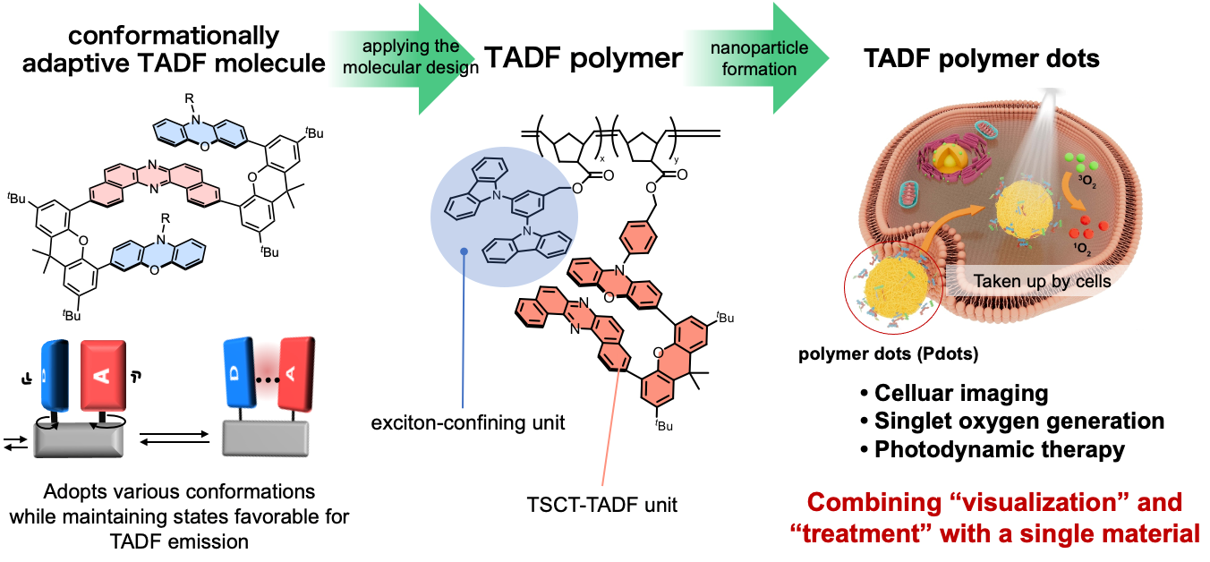
Fig. 1
Schematic overview of this study.
Original content, Credit must be given to the creator, Youhei Takeda
A research team led by the University of Osaka has developed a single synthetic material that allows imaging of cells and has the potential to cause cancer cell death when exposed to light
Osaka, Japan – A research team led by Professor Youhei Takeda at the University of Osaka, in collaboration with Professor Zachary M. Hudson at the University of British Columbia (UBC), Canada, has developed a polymer material that combines fluorescent cell imaging with the potential to kill cancer cells when activated by light. The material could offer a simpler approach to developing “nanotheranostic” technologies, which bring disease imaging and treatment together in a single platform.
Nanotheranostic materials are challenging to develop, because one material must accommodate the different demands of both imaging and therapy. In their study published in Chem & Bio Engineering, the research team formed their polymer into nanoparticles known as polymer dots, creating structures that can emit fluorescent light for imaging while also generating singlet oxygen, a reactive form of oxygen that can damage cells.
The material uses a phenomenon called “thermally activated delayed fluorescence” (TADF) to produce orange fluorescence. Because this delayed fluorescence lasts much longer than the natural background glow of cells and tissue, it could enable clearer, higher-contrast imaging in the future. Under suitable light-irradiation conditions, it can also generate singlet oxygen, providing the potential for “photodynamic therapy,” a technique that uses light-activated chemical processes to damage targeted cells.
The distinctive properties of this polymer arise in part from “through-space charge transfer,” in which electronic interactions occur between molecular groups that are stacked closely together in space, rather than through the chemical bonds that link them. This polymer also exhibits conformational adaptability, allowing its molecular structure to adopt different arrangements while retaining its functional properties. Together, these features prevent the loss of light emission that typically occurs when light-emitting molecules are crowded together.
Unlike some approaches that combine imaging and therapy by incorporating a separate light-activated compound (a photosensitizer), this material integrates both functions within a single platform. This could simplify the design process for future applications linking imaging and therapy.
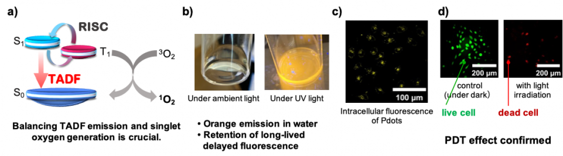
“An important challenge when designing these materials is maintaining their properties in water, which mimics the environment in the body. Often, light-emitting molecules lose their glow when packed closely together in nanoparticles. Our experiments revealed that this material retained its orange emission and long-lived delayed fluorescence in water, even with a high proportion of light-emitting units,” explains lead author Nae Aota. “Through the JST-ASPIRE program, I had the opportunity to conduct research at the University of British Columbia in Canada. This experience was invaluable in broadening my scientific perspective and strengthening our international research collaboration.”
Laboratory experiments showed that the polymer dots were taken up by cells and accumulated predominantly in lysosomes, compartments within cells involved in processing and recycling cellular material. The fluorescence of the polymer enabled the cells to be visualized, while light irradiation triggered singlet oxygen generation, demonstrating the potential of this material for photodynamic therapy.
“Thus, we have developed a novel TADF polymer material that can integrate the functions of cellular visualization and light-activated therapy within a single platform,” explains senior author Youhei Takeda. “This achievement also highlights the importance of international collaboration, particularly our partnership with UBC, supported by JST's ASPIRE program. We hope that this collaboration will accelerate the development of advanced luminescent materials for future biomedical applications.”
The findings provide a foundation for further research into multifunctional materials for bioimaging and photodynamic therapy, including their potential application for treating diseases such as cancer.
###
The article, “Conformationally Adaptive Through-Space Charge Transfer TADF Polymer Dots for Bioimaging and Photodynamic Therapy,” will be published in Chem & Bio Engineering at DOI: https://doi.org/10.1021/cbe.6c00141
Fig. 2
a) Relationship between TADF emission and singlet oxygen generation; b) photographs of Pdots dispersed in water under ambient light and UV irradiation; c) fluorescence imaging of Pdots in cells; and d) photodynamic therapy (PDT) effect upon light irradiation.
Original content, Credit must be given to the creator, Youhei Takeda
About The University of Osaka
The University of Osaka was founded in 1931 as one of the seven imperial universities of Japan and is now one of Japan's leading comprehensive universities with a broad disciplinary spectrum. This strength is coupled with a singular drive for innovation that extends throughout the scientific process, from fundamental research to the creation of applied technology with positive economic impacts. Its commitment to innovation has been recognized in Japan and around the world. Now, The University of Osaka is leveraging its role as a Designated National University Corporation selected by the Ministry of Education, Culture, Sports, Science and Technology to contribute to innovation for human welfare, sustainable development of society, and social transformation.
Website: https://resou.osaka-u.ac.jp/en
Advertisement