

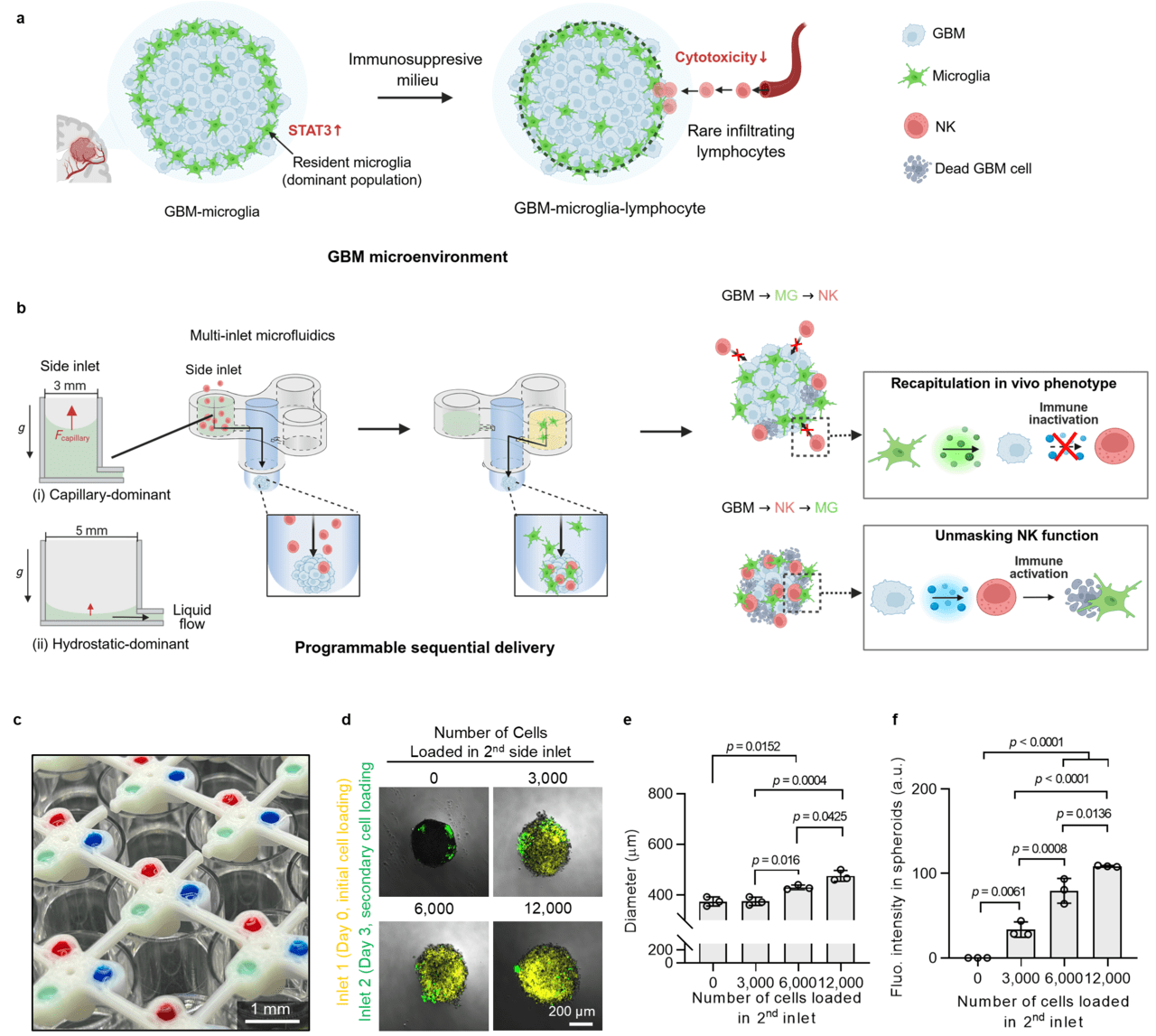
Figure 1. Sequential immunoregulation in glioblastoma (GBM) is not fully resolved in vivo but is controllable in vitro.
(A) GBM tumors in vivo are characterized by resident MG dominance and reduced natural killer (NK) cell cytotoxicity. While this outcome is evident, the temporal order and causal link between early MG activity and subsequent NK suppression cannot be directly observed. (B) Programmable delivery of immune cells through a multi-inlet microfluidic device recapitulates the suppressed phenotype observed in vivo, whereas reversing the order unmasks NK function, revealing IL12A induction and enhanced cytotoxicity. (C) Images showing spatial fluid delivery using dyed solutions injected from the 3 inlets of the multi-inlet microfluidic device. Scale bar: 1 mm. (D) Photograph showing the incorporation of cells injected through the 2nd (side) inlet. Scale bar: 200 μm. (E) Measurement of spheroid diameter as a function of the number of cells injected from the 2nd inlet. n = 3. (F) Quantitative analysis of fluorescence intensity in the spheroid region corresponding to cells delivered from the 2nd inlet. n = 3. Statistical analysis was done by 1-way ANOVA followed by Tukey’s post hoc test.
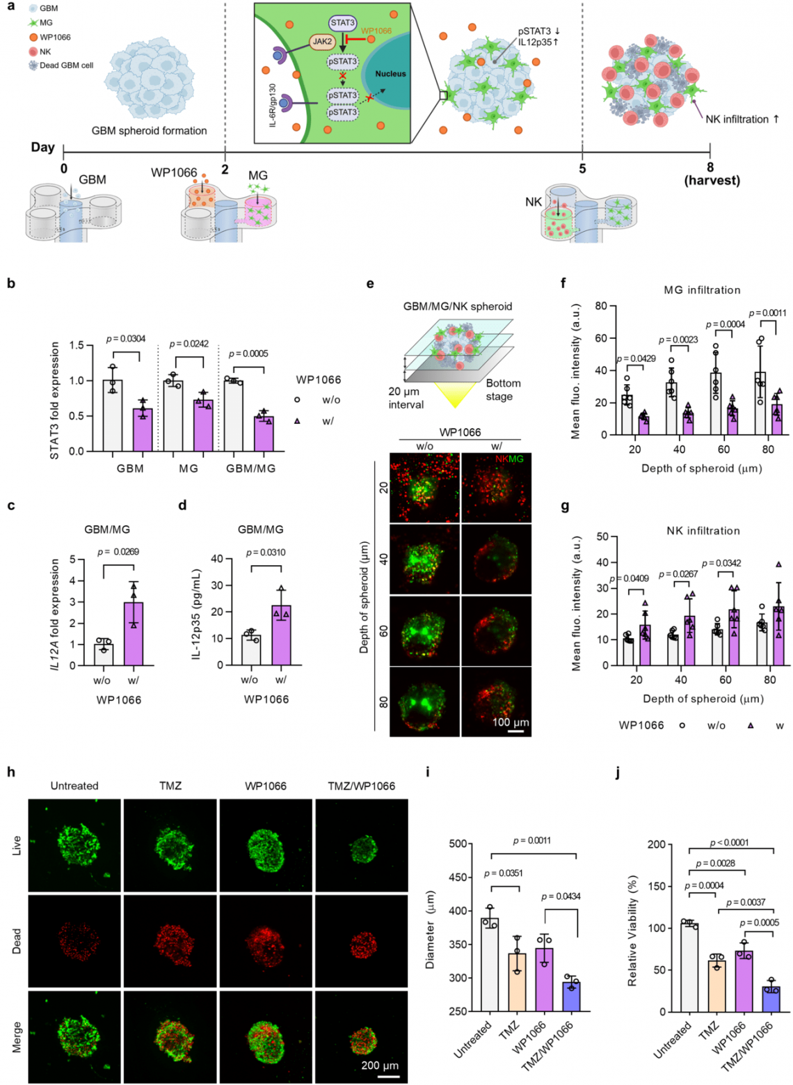
Figure 2. STAT3 inhibition restores IL12A expression and enhances natural killer (NK) infiltration, sensitizing glioblastoma (GBM) spheroids to TMZ.
(A) Schematic of experimental timeline for WP1066 and NK cell treatment in GBM/MG spheroids.
(B) STAT3 mRNA expression in GBM, MG, and GBM/MG spheroids with or without WP1066 treatment. n = 3. (C and D) IL12A mRNA expression (C) and release of IL-12p35 (D) in GBM/MG spheroids with or without WP1066 treatment. n = 3. (E) Representative schematic and confocal z-stack images (20-μm intervals) of GBM/MG/NK spheroids treated with or without WP1066, showing NK and MG distribution at different spheroid depths. Scale bar: 100 μm. (F and G) Quantification of MG (F) and NK (G) infiltration intensities across spheroids. n = 6. (H) Live/dead staining of GBM/MG/NK spheroids under different treatments: untreated, TMZ, WP1066, and TMZ/WP1066. Scale bar: 200 μm. (I and J) Quantification of spheroid diameter (I) and relative viability (J) under different treatment conditions. n = 3. Statistical significance was determined using an unpaired 2-tailed t test (B-D, F, G) and 1-way ANOVA with Tukey’s post hoc test (I and J).
A joint research team led by Professor Sungsu Park of Sungkyunkwan University and Professor Sun-Ha Paek of Seoul National University College of Medicine has demonstrated for the first time that the sequence in which immune cells interact with cancer cells can critically influence the therapeutic response in glioblastoma. The findings reveal the importance of timing in tumor–immune interactions and suggest a new strategy for optimizing personalized cancer immunotherapy.
Glioblastoma (GBM), one of the most lethal brain tumors, is characterized by a strongly immunosuppressive microenvironment driven in part by the extensive infiltration of microglia, the brain’s resident immune cells.
Consequently, natural killer (NK) cells, which directly attack cancer cells, have limited ability to penetrate the tumor, reducing their therapeutic effectiveness. Conventional static cell culture models also have limitations in capturing the dynamic, time-dependent interactions between immune cells and tumor tissue.
To investigate these time-dependent interactions, the team developed a multi-inlet microfluidic platform that enables precise control over the timing and sequence of cell introduction in a 3D tumor model. Using the platform, the researchers compared identical combinations of cancer cells, microglia, and NK cells while varying only the order in which the immune cells were introduced.
The results showed that when microglia reached the cancer cells first, STAT3 signaling was activated, limiting NK-cell infiltration and promoting tumor growth. In contrast, when NK cells reached the cancer cells first, IL12A, a key immune-activating factor, was strongly upregulated in the cancer cells, leading to sustained tumor suppression. These findings demonstrate that even with the same cellular composition, the tumor immune response can differ markedly depending on which immune cells interact with the cancer cells first. Furthermore, the team demonstrated the translational potential of the platform using patient-derived glioblastoma cells.
When the STAT3 inhibitor WP1066 was combined with temozolomide (TMZ), NK-cell infiltration into the tumor significantly increased. The combination also enhanced tumor cell death, demonstrating a synergistic therapeutic effect.
The study lays the groundwork for a patient-tailored precision medicine platform. By recreating a patient’s tumor immune microenvironment on a chip, the platform could help identify effective drug combinations and optimal treatment timing. The core technology has been commercialized by ORGANOPlus Inc. (https://organoplus.kr) as CANORGAN and is now available for research use. Professor Sungsu Park said, “I am pleased that we were able to elucidate the temporal interactions between immune cells within tumor tissue using engineering technology—interactions that have been difficult to observe using animal models or conventional in vitro culture. “We will continue to develop this technology as a patient-tailored drug screening platform for various refractory solid cancers.”
This work was supported by the Ministry of Science and ICT and the National Research Foundation of Korea (NRF). The findings were published in Neuro-Oncology, a leading international journal in the field of brain tumor research.