


A team of researchers at the University of Osaka has revealed the stepwise mechanism by which bacteria assemble a molecular bushing around the drive shaft of the flagellar motor. The ring first forms while tightly bound to the shaft and is then released from its surface, allowing it to support the high-speed, low-friction rotation required for bacterial movement. By clarifying how a complex biological machine can assemble itself with remarkable precision, this discovery provides valuable design principles for artificial nanomachines and may contribute to future technologies such as efficient drug-delivery systems and other biomedical devices.
Fig .1
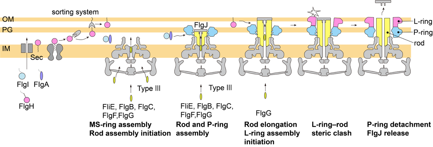
The LP-ring assembly process around the rod
FlgI monomers assemble around the rod to form the P-ring, which is firmly attached to the rod surface with its slightly elliptic shape and small tilt. FlgH monomers are further translocated to the outer membrane (OM) and assemble on the P-ring to form the L-ring after the rod grows and penetrates the peptidoglycan layer (PG). The L-ring formed on the tilted elliptic P-ring clashes with the rod surface, forcing the P-ring to be oriented perpendicular to the rod axis and dissociate from the rod surface so that the LP-ring becomes a non-elliptic cylinder surrounding the free-standing rod to be a functional bushing.
©Original content, Credit must be given to the creator., Adapted from Figure 5 of "Structural insights into the assembly mechanism of the molecular bushing in the bacterial flagellar motor" by Tomoko Yamaguchi et al., 2026, Communications Biology
Structural rearrangements enable precise positioning, release, and low-friction rotation
Osaka. Japan - The bacterial flagellum is a motility organelle that enables bacteria to swim and is composed of more than 30 different proteins. Researchers have long sought to understand how this complex nanomachine self-assembles from many parts in the correct order and at the correct positions. In a study published in Communications Biology, a research team at the University of Osaka revealed the mechanism by which the flagellar motor assembles its bushing around the drive shaft.
The LP-ring acts as a molecular bushing surrounding the drive shaft called the rod and supports high-speed rotation of the rod at low friction. During motor assembly, the rod first polymerizes, after which the P-ring forms around a specific part of the rod. The L-ring subsequently assembles on top of the P-ring, and the completed LP-ring is released from the rod surface to become a bushing.
“Because the rod rotates within the mature LP-ring without physical contact, it has long been unclear how the LP-ring is accurately positioned during assembly and how it is later released from the rod,” says Tomoko Yamaguchi. “To understand how the LP-ring assembles, we needed structural information on the assembly intermediate. “
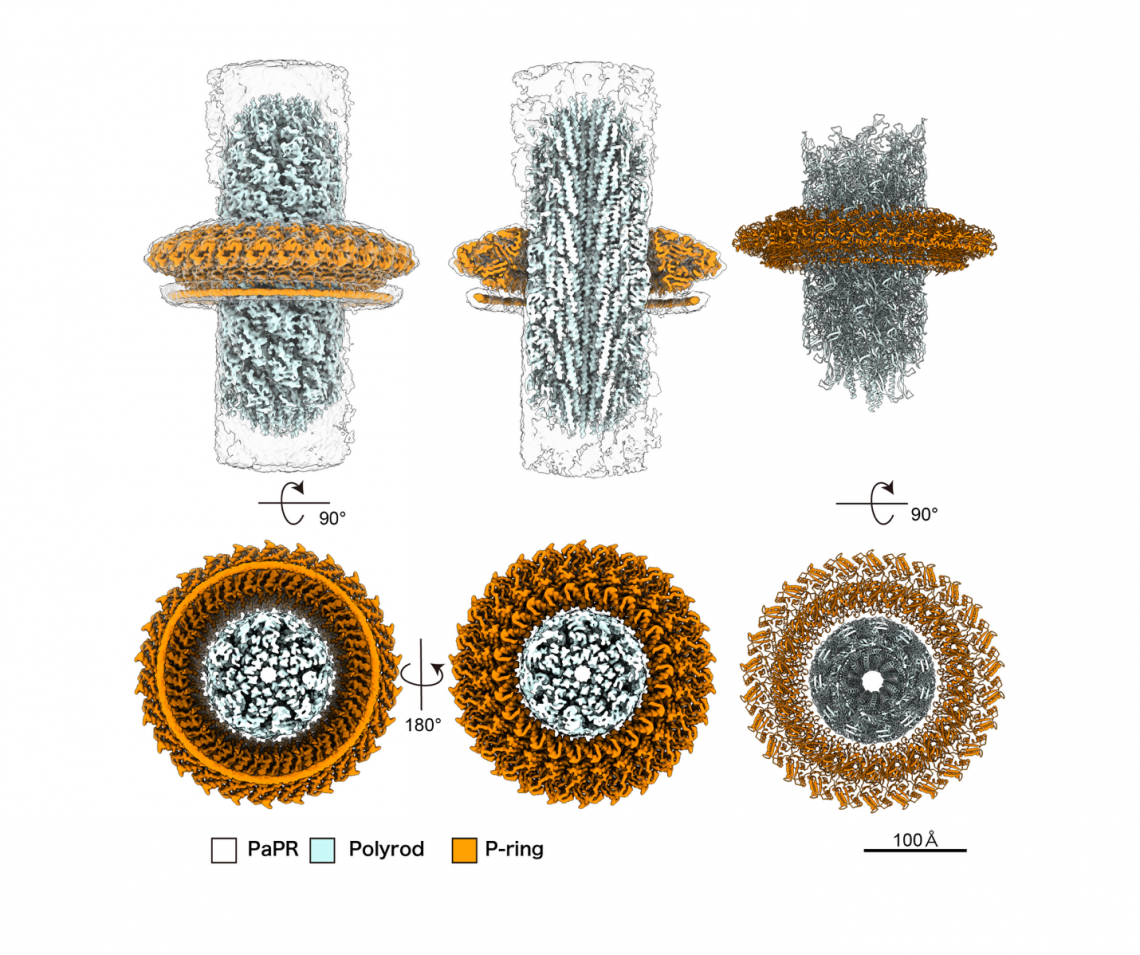
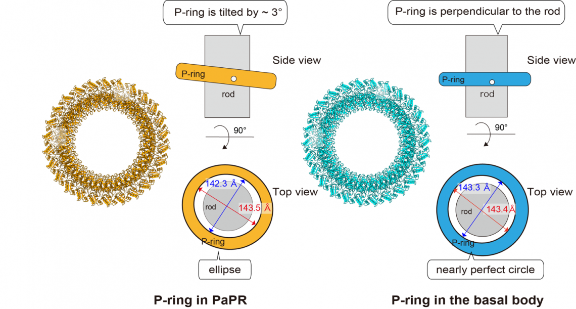
To capture the assembly intermediate, the researchers analyzed a polyrod–P-ring complex (PaPR), in which multiple P-rings assemble around genetically-modified elongated rods. Using electron cryomicroscopy (cryoEM) single particle analysis, they found that the P-ring initially adopts a slightly elliptical and tilted conformation to increase the contact area with the rod surface to bind to a specific position of the rod. Then, upon formation of the L-ring, steric hindrance arises between the rod and the L-ring, causing the entire LP-ring to detach from the rod surface and become a functional bushing.
Fig. 2
Overall structure of the PaPR complex
The cryoEM density maps of the PaPR complex (left and center) and the corresponding atomic models (right).
©CC BY, 2026, Tomoko Yamaguchi et al., Structural insights into the assembly mechanism of the molecular bushing in the bacterial flagellar motor, Communications Biology
“Artificially reproducing the autonomous assembly of a complex biomolecular machine remains major technological challenge,” says the senior author Keiichi Namba. “The bacterial flagellum achieves this process with remarkable precision through self-organization.”
The newly identified mechanism for constructing and releasing a molecular bearing provides important design principles for self-assembling nanomachines and could contribute to future nanotechnology applications, including highly efficient drug delivery systems (DDSs).
###
The article, “Structural insights into the assembly mechanism of the molecular bushing in the bacterial flagellar motor,” was published in Communications Biology at DOI: https://doi.org/10.1038/s42003-026-10640-y
Fig. 3
Comparison of the P-ring structures in the PaPR and basal body.
The P-ring in the PaPR (assembly intermediate) is tilted by approximately 3º relative to the rod axis, whereas the P-ring in the basal body (matured form) is perpendiculer to the rod axis. The red and blue double-sided arrows are the major and minor axes of the fitted ellipses, respectively. The P-ring in the PaPR is slightly elliptical, whereas that of the basal body is a nearly perfect circle.
©Original content, Credit must be given to the creator., Adapted from Figure 2c of "Structural insights into the assembly mechanism of the molecular bushing in the bacterial flagellar motor" by Tomoko Yamaguchi et al., 2026, Communications Biology
About The University of Osaka
The University of Osaka was founded in 1931 as one of the seven imperial universities of Japan and is now one of Japan's leading comprehensive universities with a broad disciplinary spectrum. This strength is coupled with a singular drive for innovation that extends throughout the scientific process, from fundamental research to the creation of applied technology with positive economic impacts. Its commitment to innovation has been recognized in Japan and around the world. Now, The University of Osaka is leveraging its role as a Designated National University Corporation selected by the Ministry of Education, Culture, Sports, Science and Technology to contribute to innovation for human welfare, sustainable development of society, and social transformation.
Website: https://resou.osaka-u.ac.jp/en
Advertisement