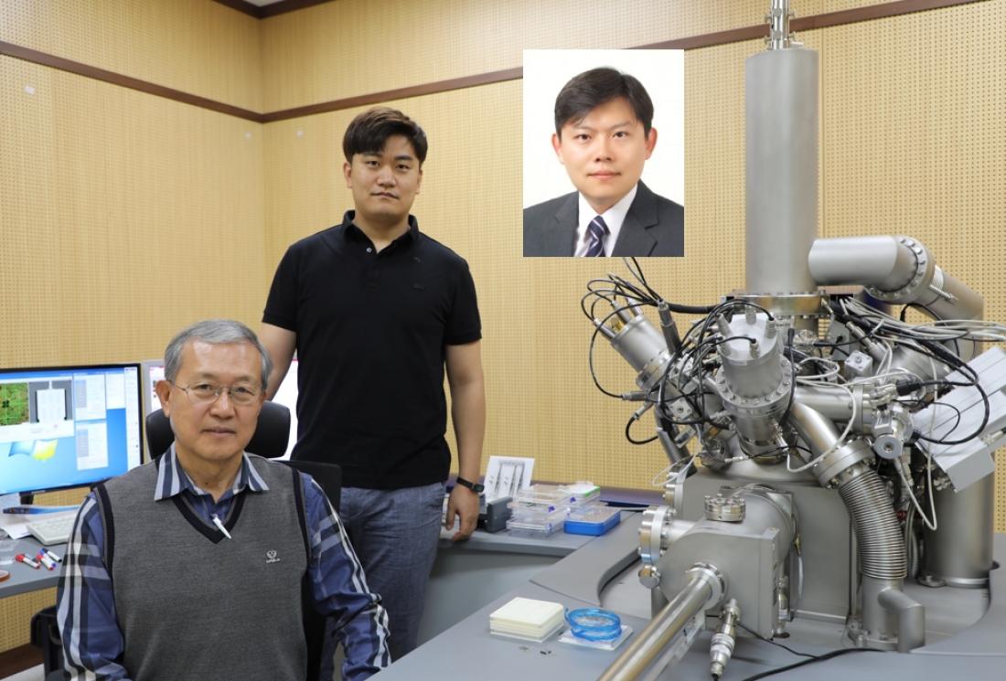
Photo1.(From left to right) Professor Dae Won Moon, Mr. Young-Ho Park., and Professor Su-Il In
A multitude of proteins are involved in degenerative diseases, and Alzheimer’s disease is no exception. The proposed technique could deepen our understanding of how anomalous protein interactions cause such diseases, which is crucial to develop early detection protocols and treatments.
Degenerative diseases such as Alzheimer’s often involve complex interactions between multiple proteins and other biomolecules. Understanding these interactions using existing imaging technologies is difficult because of insufficient resolution and the impossibility of simultaneously detecting many different proteins.
In a recent interdisciplinary study, a research team led by Professors Dae Won Moon and Su-Il In from Daegu Gyeongbuk Institute of Science and Technology developed an innovative approach that extends the applications of the technique ‘secondary-ion mass spectrometry’ from its original purpose in the semiconductor industry to the biomedical imaging field. Secondary-ion mass spectrometry allows one to analyze the composition of surfaces and offers very high resolution. Using this technology to image proteins was impossible—until now.
In this novel approach, different metal oxide nanoparticles are individually attached to antibodies that bind to specific target proteins. The mass spectrometer can easily detect these nanoparticles even at low irradiation doses, which leaves the cell tissue intact and allows for multiple analyses on the same cells. Through this approach, it is theoretically possible to analyze tens of proteins simultaneously. This vastly surpasses existing fluorescence-based approaches, which allow for the simultaneous imaging of around four proteins.
The researchers used their method to compare the distribution of proteins in the brain tissue of mice that were either healthy or had Alzheimer’s. They showed that valuable insight could be gained from observing multiplex protein distributions in the ‘hippocampus’ section of the brain and demonstrated how these differed between healthy and diseased mice.
Achieving these results required combining knowledge from many disciplines. Prof. Moon remarks, “For me, a special highlight of this work is the collaboration of many researchers from different backgrounds, such as chemists, nano-particle experts, medical doctors, and biologists. It was not easy and took a long time, but it was exciting to see the progress. We can now visualize multiple proteins on cell membranes with a spatial resolution of 300 nanometers.” He expects this new imaging approach to become an important tool to deepen our understanding on degenerative diseases. “The proposed technique could be used to figure out the early stages of Alzheimer’s disease, in turn allowing for early detection and treatment,” he states, hopefully.
Reference
|
Authors: |
Dae Won Moon*1, Young Ho Park2, Sun Young Lee1, Heejin Lim1, SuHwa Kwak3, Minseok S. Kim1, Hyunmin Kim4, Eunjoo Kim4, Yebin Jung5, Hyang-Sook Hoe6, Sungjee Kim5, Dong-Kwon Lim7, Chul-Hoon Kim8, and Su-Il In*2 |
|
Title of original paper: |
Multiplex Protein Imaging with Secondary Ion Mass Spectrometry Using Metal Oxide Nanoparticle-Conjugated Antibodies |
|
Journal: |
ACS Applied Materials & Interfaces |
|
DOI: |
|
|
Affiliations: |
1Department of New Biology, DGIST 2Department of Energy Science and Engineering, DGIST 3Department of Computer Science and Engineering, POSTECH 4Companion Diagnostics and Medical Technology Research Group, DGIST 5Department of Chemistry, POSTECH 6Department of Neural Development and Disease, Korea Brain Research Institute (KBRI) 7KU-KIST Graduate School of Converging Science and Technology, Korea University 8Department of Pharmacology, Yonsei University College of Medicine
|
*Corresponding authors’ emails: [email protected] (Dae Won Moon), [email protected] (Su-Il In)
About Daegu Gyeongbuk Institute of Science and Technology (DGIST)
Daegu Gyeongbuk Institute of Science and Technology (DGIST) is a well-known and respected research institute located in Daegu, Republic of Korea. Established in 2004 by the Korean Government, the main aim of DGIST is to promote national science and technology, as well as to boost the local economy.
With a vision of “Changing the world through convergence", DGIST has undertaken a wide range of research in various fields of science and technology. DGIST has embraced a multidisciplinary approach to research and undertaken intensive studies in some of today's most vital fields. DGIST also has state-of-the-art-infrastructure to enable cutting-edge research in materials science, robotics, cognitive sciences, and communication engineering.
Website: https://www.dgist.ac.kr/en/html/sub01/010204.html
About the author
D. W. Moon, first author and co-corresponding author of this work, has studied surface analytical tools, such as SIMS and MEIS, for semiconductor industries for 40 years. These techniques employ ion beams to analyze the surfaces and interfaces of semiconductor ultrathin films. For the past 20 years, he has been working on how to apply nano-analysis tools developed for use in an ultrahigh vacuum as bioimaging tools for materials and particularly cells in liquids.


