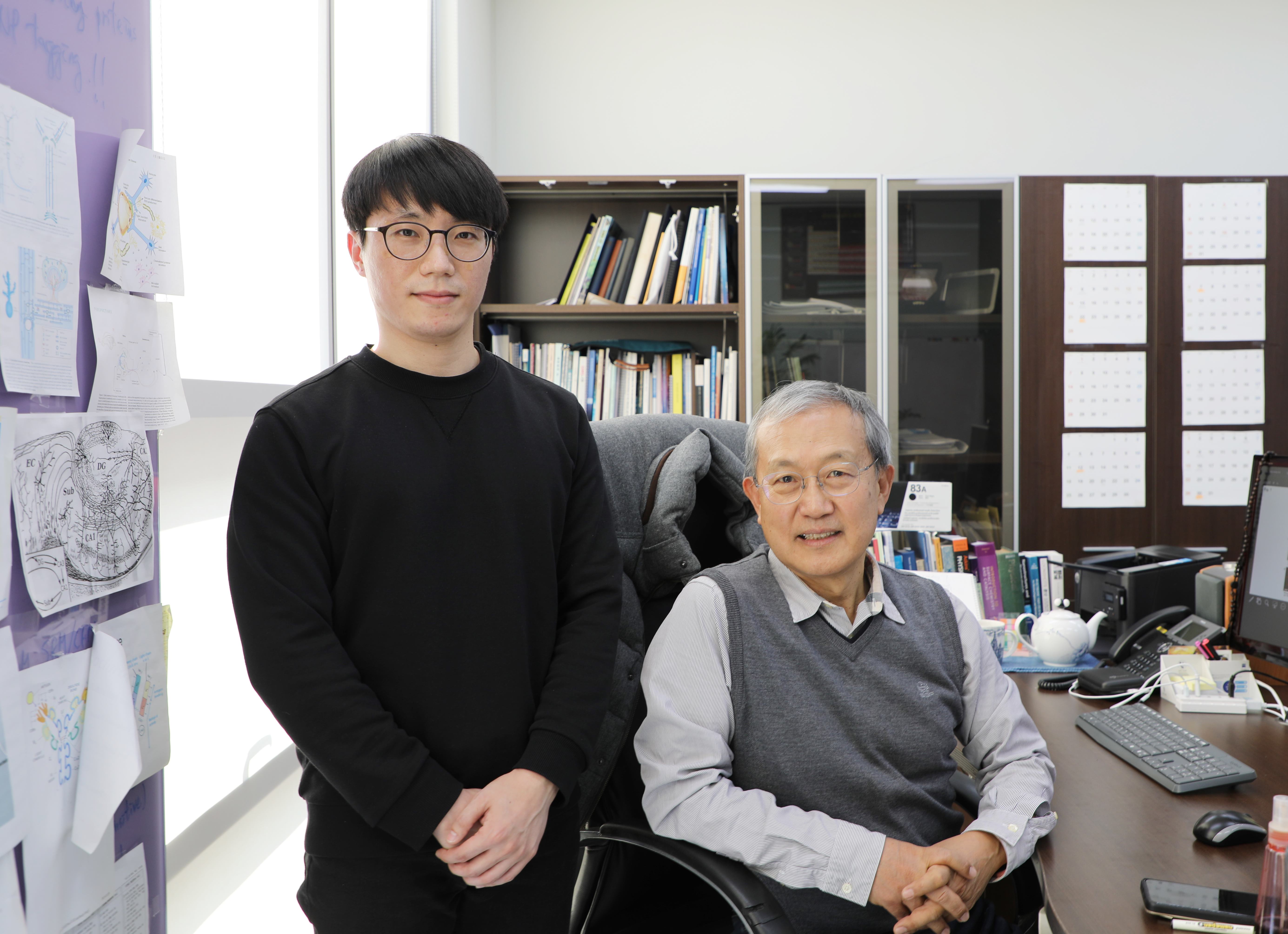
Professor Dae Won Moon (sitting) and Dr. Heejin Lim (standing) in their lab at DGIST, Korea.
Understanding in detail the molecular structure of the cell membrane is important for learning more about the underlying cellular mechanisms of diseases. New techniques are needed to look at these miniscule structures in high resolution and with high accuracy
Scientists graphene coat cells to keep them alive and wet even in vacuum, significantly advancing the field of bioimaging
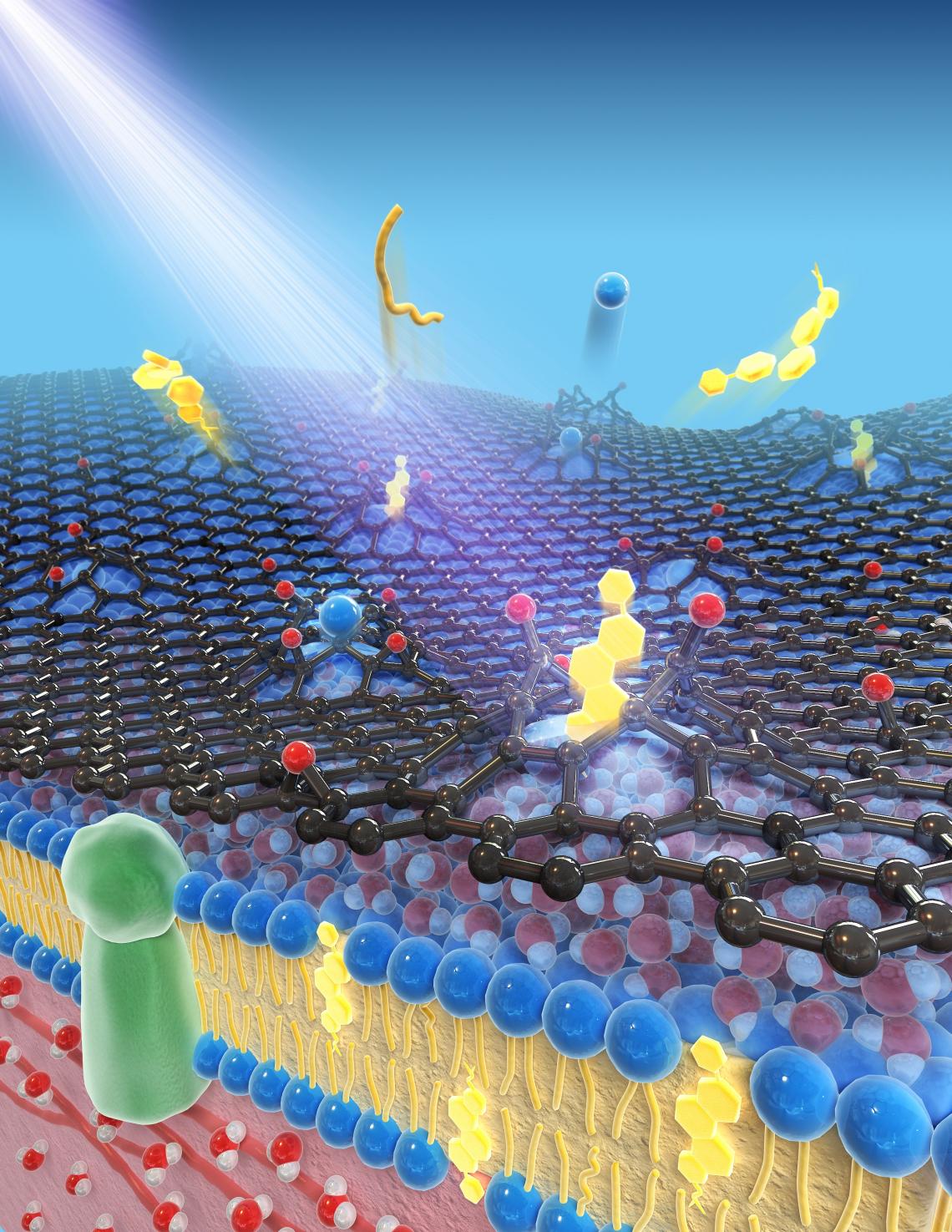
Researchers from DGIST have now found a way to keep living, wet cells viable in an ultra-high-vacuum environment, using graphene, allowing—like never before—accurate high-resolution visualization of the undistorted molecular structure and distribution of lipids in cell membranes. This could enhance our bioimaging abilities considerably, improving our understanding of mechanisms underlying complex diseases such as cancers and Alzheimer’s.
With every passing day, human technology becomes more refined and we become slightly better equipped to look deeper into biological processes and molecular and cellular structures, thereby gaining greater understanding of mechanisms underlying diseases such as cancer, Alzheimer’s, and others.
Today, nanoimaging, one such cutting-edge technology, is widely used to structurally characterize subcellular components and cellular molecules such as cholesterol and fatty acids. But it is not without its limitations, as Professor Dae Won Moon of Daegu Gyeongbuk Institute of Technology (DGIST), Korea, lead scientist in a recent groundbreaking study advancing the field, explains: “Most advanced nanoimaging techniques use accelerated electron or ion beams in ultra-high-vacuum environments. To introduce cells into such an environment, one must chemically fix and physically freeze or dry them. But such processes deteriorate the cells’ original molecular composition and distribution.”
Prof. Moon and his team wanted to find a way to avoid this deterioration. “We wanted to apply advanced nanoimaging techniques in ultra-high-vacuum environments to living cells in solution without any chemical and physical treatment, not even fluorescence staining, to obtain intrinsic biomolecular information that is impossible to obtain using conventional bioimaging techniques,” Dr. Heejin Lim, a key member of the research team, explains. Their novel solution is published in Nature Methods.
Their technique involves placing wet cells on a collagen-coated wet substrate with microholes, which in turn is on top of a cell culture medium reservoir. The cells are then covered with a single layer of graphene. It is the graphene that is expected to protect the cells from desiccation and cell membranes from degradation.
Through optical microscopy, the scientists confirmed that, when prepared this way, the cells remain viable and alive up to ten minutes after placing in an ultra-high-vacuum environment. The scientists also performed nanoimaging, specifically, secondary ion mass spectrometry imaging, in this environment for up to 30 mins. The images they captured within the first ten minutes paint a highly detailed (submicrometer) picture of the true intrinsic distribution of lipids in their native states in the cell membranes; for this duration, the membranes underwent no significant distortion.
With this method too, however, a cascade of ion beam collisions at a point on the graphene film can create a big enough hole for some of the lipid particles to escape. But while this degradation to the cell membrane does occur, it is not significant within the ten-minute window and there is no solution leakage. Further, the graphene molecules react with water molecules to self-repair. So, overall, this is a great way to learn about cell membrane molecules in their native state in high resolution.
“I imagine that our innovative technique can be widely used by many biomedical imaging laboratories for more reliable bioanalyses of cells and eventually for overcoming complex diseases,” says Prof. Moon.
Will this innovation become the norm? Only time will tell!
Reference
|
Authors: |
Heejin Lim1, Sun Young Lee1, Yereum Park2, Hyeonggyu Jin3, Daeha Seo3, Yun Hee Jang2 and Dae Won Moon1,4 |
|
Title of original paper: |
Mass spectrometry imaging of untreated wet cell membranes in solution using single-layer graphene |
|
Journal: |
Nature Methods |
|
DOI: |
|
|
Affiliations: |
1Department of New Biology, DGIST, Daegu, Republic of Korea 2Department of Energy Science and Engineering, DGIST, Daegu, Republic of Korea 3Department of Emerging Materials Science, DGIST, Daegu, Republic of Korea 4School of Undergraduate Studies, DGIST, Daegu, Republic of Korea |
*Corresponding author’s email: [email protected]
About Daegu Gyeongbuk Institute of Science and Technology (DGIST)
Daegu Gyeongbuk Institute of Science and Technology (DGIST) is a well-known and respected research institute located in Daegu, Republic of Korea. Established in 2004 by the Korean Government, the main aim of DGIST is to promote national science and technology, as well as to boost the local economy.
With a vision of “Changing the world through convergence", DGIST has undertaken a wide range of research in various fields of science and technology. DGIST has embraced a multidisciplinary approach to research and undertaken intensive studies in some of today's most vital fields. DGIST also has state-of-the-art-infrastructure to enable cutting-edge research in materials science, robotics, cognitive sciences, and communication engineering.
Website: https://www.dgist.ac.kr/en/html/sub01/010204.html
About the authors
Dr. Dae Won Moon is Professor at the Department of New Biology and is affiliated to the NanoBio Imaging Laboratory at DGIST. He began his career with a PhD in Chemistry from Pennsylvania State University, USA, in 1984, and is now on the editorial board of the journal Critical Reviews in Solid State and Materials Science. His research interests include coherent Raman scattering, surface plasmon resonance imaging ellipsometry, imaging mass spectrometry, and time-of-flight medium energy ion scattering. For his exceptional contribution to the field, including over 180 publications, he has won several awards in Korea.