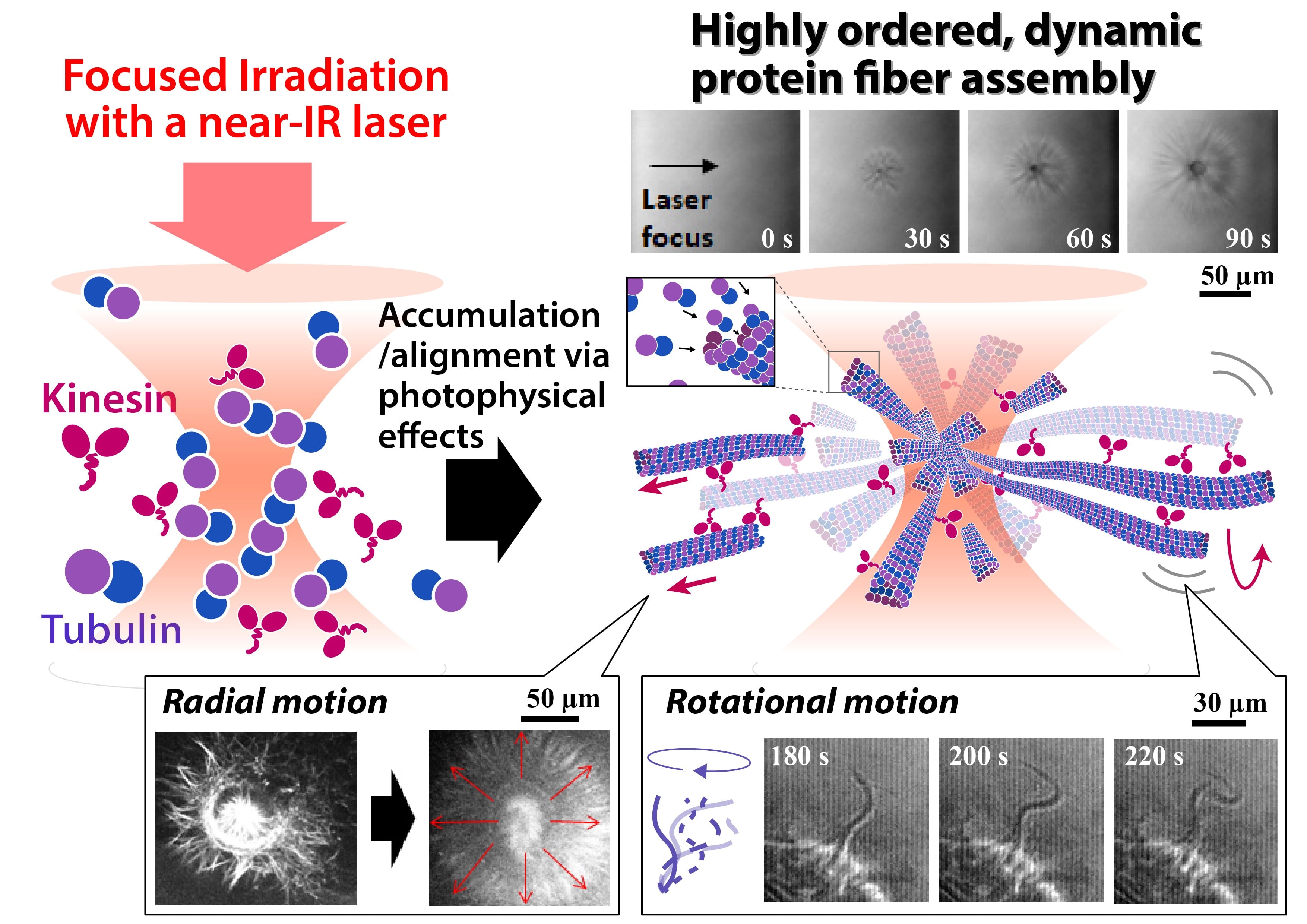
Fig. 1
Tightly focused laser irradiation can accumulate tubulin proteins at/around the laser focus, which leads to the formation of highly ordered microtubule assemblies. The assemblies can exhibit various dynamic behaviors such as radial motion, bundling, and flagella-like rotation with motor protein and chemical energy, highlighting as a unique tool for spatiotemporal control of protein assembly without chemical/biological modifications.
Researchers use the force of a focused laser beam to make precise models of cytoskeletons in living cells
Osaka, Japan – Networks of protein fibers play important roles in living cells. To understand the dynamical behavior of these networks, model networks are needed to perform in vitro studies. However, fabrication of protein networks similar to those in cells has proved difficult, as current methods could affect the biological function of these proteins – ultimately impacting our understanding of any findings.
Now, researchers at The University of Osaka and Saitama University have used a laser beam to precisely fabricate a network of protein fibers. This exciting discovery was recently reported in Advanced Science.
The shape of living cells is determined by an internal network of protein fibers called a cytoskeleton. The cytoskeletal structure is dynamic, as the key nodes for cell function shift over time. One such cell function can be witnessed with motor proteins, which convert chemical energy into mechanical work. These proteins walk along cytoskeletal tracks to drive muscle contraction and transport components across the cell.
In vitro models have been constructed to understand the structure–motion relationship of cytoskeletal networks, meaning how the organization of molecules affects its dynamics. Some models have been formed via self-organization, which is the spontaneous arising of structures through interactions between protein fibers.
However, the arrangement and evolution of the network structure cannot be controlled using self-organization alone. To tackle this, ultraviolet or blue light has been employed in research to spark the formation of a protein network at specific locations.
The native biomolecules forming the network need to be chemically modified to respond to light, which could affect their biological functions and, crucially, any resulting conclusions. Light triggers organization effectively, but can cause interference when fluorescence imaging is used for visualization.
To circumvent these problems, the team tested whether the photophysical effects of a focused laser beam could be used to fabricate a fibrous protein network in a non-invasive manner.
“The electric field of the focused laser beam generated an optical force on very small protein molecules,” explains lead author, Hiroshi Yoshikawa. “The optical force caused these molecules to accumulate at the laser focus without bringing them into physical contact. As a result, highly ordered arrangements of protein-fiber networks formed.”
The fabricated networks exhibited various dynamic motions similar to those observed in living cells, such as translational motion and flagella-like rotation.
“Our approach does not require chemical modification of molecules, reducing the likelihood that native biomolecules lose their biological functions”,” reports Hiroshi Yoshikawa. “Additionally, the laser wavelength used is in the near-infrared regime, which does not overlap with the wavelengths of light sources used for fluorescence imaging.”
This technology could be applied in biological fields to determine mechanisms involving the cytoskeleton, such as cell division, migration, and adhesion. Interestingly, the technology could also have applications beyond biology, including creating protein-based actuators (robotic “muscles”).
###
The article, “Spatiotemporal Control of Formation of Dynamic Protein Fiber Assemblies via Photophysical Effects of a Focused Laser Beam,” was published in Advanced Science at DOI: https://doi.org/10.1002/advs.75531
A higher-resolution version is available at the link below.
A higher-resolution version is available at the link below.
About The University of Osaka
The University of Osaka was founded in 1931 as one of the seven imperial universities of Japan and is now one of Japan's leading comprehensive universities with a broad disciplinary spectrum. This strength is coupled with a singular drive for innovation that extends throughout the scientific process, from fundamental research to the creation of applied technology with positive economic impacts. Its commitment to innovation has been recognized in Japan and around the world. Now, The University of Osaka is leveraging its role as a Designated National University Corporation selected by the Ministry of Education, Culture, Sports, Science and Technology to contribute to innovation for human welfare, sustainable development of society, and social transformation.
Website: https://resou.osaka-u.ac.jp/en