


For decades, scientists have understood proteins primarily through two defining features: their amino acid sequence and their three-dimensional structure. This framework has driven major advances in biology, biotechnology, and medicine, culminating in recent artificial intelligence tools capable of predicting protein structures with remarkable accuracy. Yet a fundamental challenge remains: even when a protein's sequence and structure are known, predicting its function, interactions, and behavior often remains difficult.
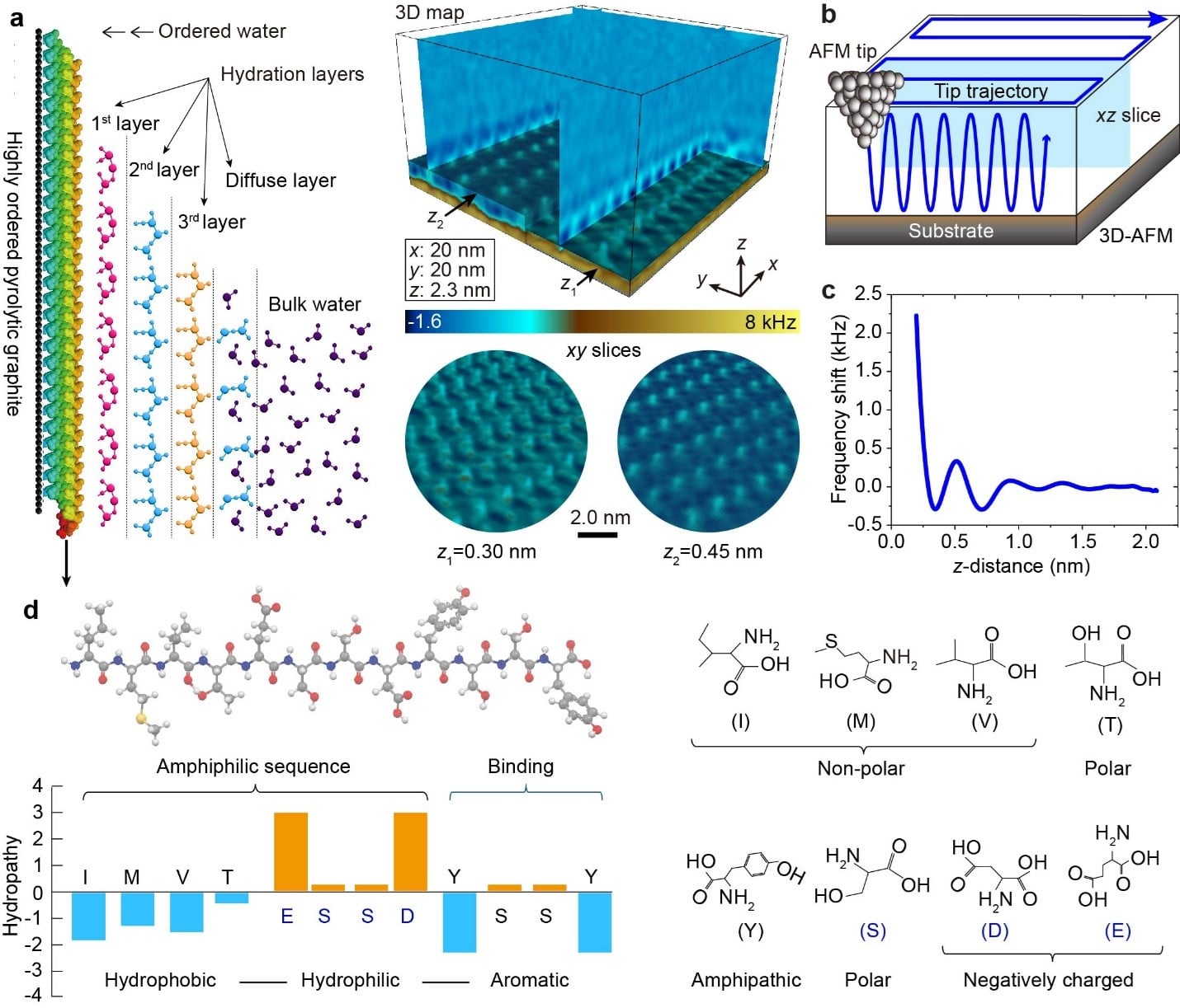
An international team of researchers from Japan, Finland, Italy, and the United States now reports evidence that part of this missing information may lie in an often-overlooked component of proteins: the highly organized water surrounding them. In a study accepted for publication in Nature Communications, the team provides the first direct three-dimensional visualization of sequence-dependent hydration architectures surrounding peptide assemblies at sub-nanometer resolution using advanced three-dimensional atomic force microscopy (3D-AFM) (Fig.1).
Figure 1. Molecular structure and 3D-AFM visualization of self-assembled dodecapeptide hydration shells. a Schematic illustration of water organization adjacent to the peptide assemblies, highlighting the formation of highly ordered hydration structures within the interfacial hydration zone. b Schematic of 3D-AFM applied to self-assembled peptide nanostructures on HOPG, showing extraction of 2D xz and xy slices from the reconstructed 3D volume map. The probe performs synchronized lateral (xy) and vertical (z) scanning, with a fast sinusoidal modulation superimposed on the z-position during image acquisition and force mapping. As the probe approaches the surface, characteristic oscillatory force profiles appear c, reflecting discrete, ordered hydration layers that are progressively penetrated near the self-assembled peptide structures. d Extended molecular structure of the GrBP5-WT peptide shown in stick–ball representation alongside its amino acid sequence. The hydropathy map reveals three chemically distinct domains within the peptide. The inset in panel a presents a 3D-AFM volumetric map showing the molecular-resolution spatial organization of interfacial water surrounding peptide assemblies, including in-plane (xy) and cross-sectional (xz) views.
Yurtsever A., et al., Nat Commun (2026)
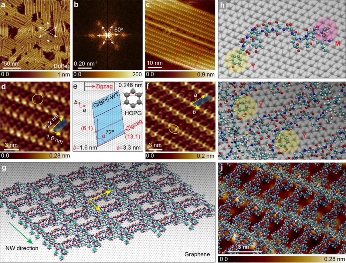
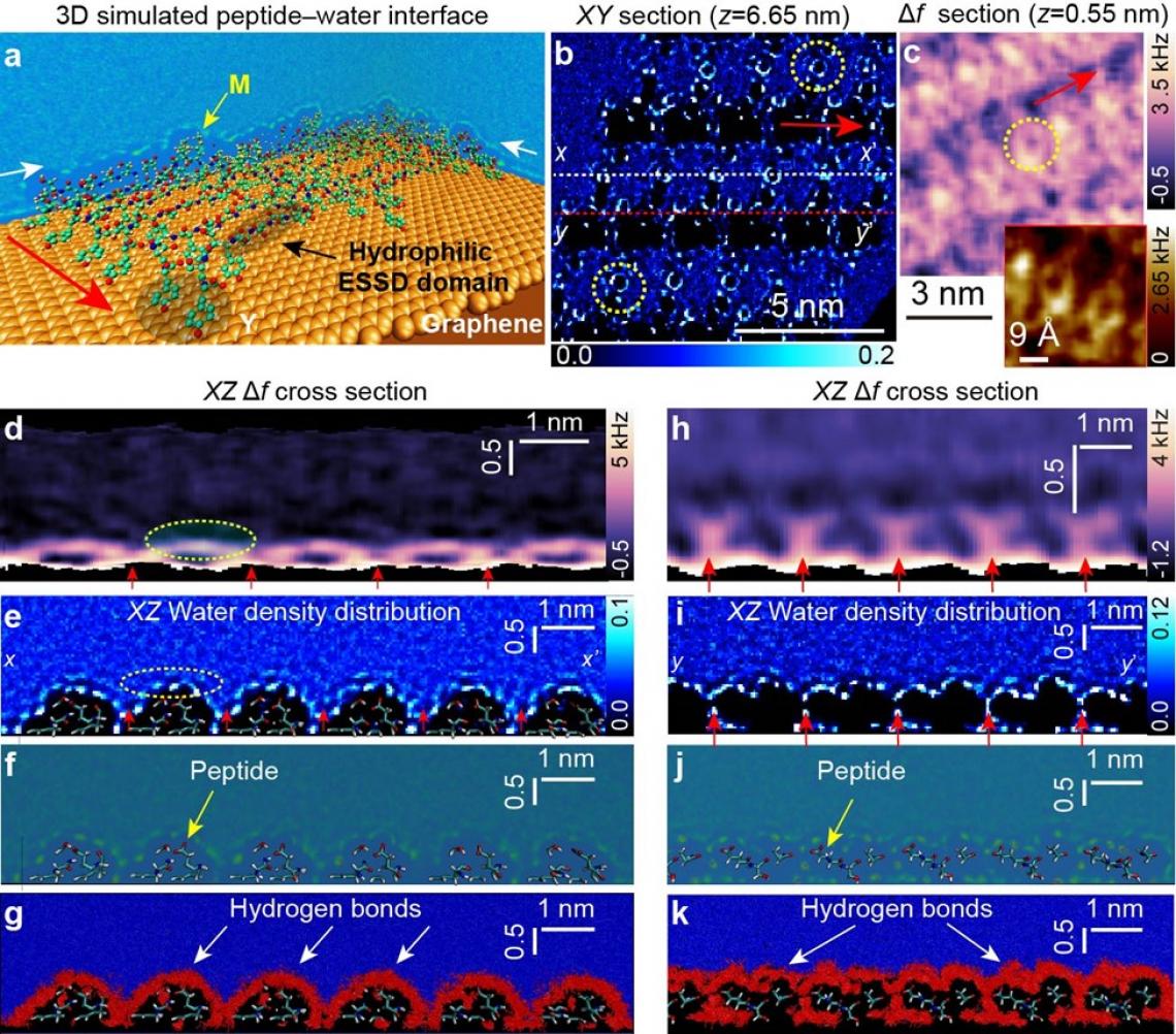
The findings reveal that water near peptide and protein surfaces is not simply a passive solvent. Instead, it forms highly organized, multilayered structures whose organization is dictated by the underlying amino acid sequence and surface chemistry (Fig.2). These hydration architectures extend multiple layers from the peptide surface and contain distinct structural signatures associated with hydrophobic, polar, aromatic, and charged amino acid residues (Fig.3).
The researchers propose that these hydration structures are not merely consequences of the protein's presence but are an integral part of protein identity and function. This has led them to introduce a new concept: the protein superstructure, defined as the protein together with its uniquely organized hydration architecture.
“Our findings suggest that proteins cannot be fully understood by sequence and structure alone,” said Prof. Mehmet Sarikaya of the University of Washington. “The highly specific hydration architecture organized by a protein appears to be part of its functional identity. We define this integrated protein-water entity as the protein superstructure.”
Water is essential to virtually every biological process. Proteins fold in water, recognize molecular partners, catalyze reactions, and function exclusively in aqueous environments. Scientists have long known that hydration influences protein folding, stability, molecular recognition, and enzymatic activity. However, directly observing the three-dimensional molecular organization of hydration water locally around proteins has remained one of the major challenges in molecular biology and biophysics. Most experimental methods provide indirect or averaged information, while computational approaches often lack direct experimental validation at molecular resolution.
Using an ordered self-assembled peptide system and high-resolution 3D-AFM, the team was able to visualize hydration structures with unprecedented detail. The results show that water organizes into sequence-specific patterns that persist across multiple hydration layers before gradually transitioning into bulk water. These patterns effectively create hydration fingerprints that reflect the underlying molecular chemistry of the peptide surface.
The discovery may help explain why predicting protein function remains challenging even when structures are well-known. Despite tremendous progress in structural biology and AI-based protein prediction, scientists still struggle to accurately predict many aspects of molecular recognition, ligand binding, enzymatic activity, and biological specificity from protein structures alone. The researchers suggest that hydration architecture may represent a previously missing layer of biological information linking sequence, structure, and function.
“A protein never exists in isolation,” said Dr. Takeshi Fukuma of Kanazawa University. “It exists together with its hydration architecture. Understanding this coupled protein-water organization may be essential for understanding how proteins function and interact in living systems.”
The implications extend across biology, medicine, biotechnology, and materials science. Incorporating hydration architecture into studies of proteins could improve understanding of protein folding, molecular recognition, and drug-target interactions. It may also provide new opportunities for protein engineering, AI-driven protein design, biomimetic materials, catalytic interfaces, biosensors, and other technologies that depend on controlling interactions in water.
Importantly, the study does not propose a new form of “biological water.” Rather, it demonstrates that interfacial water forms dynamic but persistent sequence-dependent structures imposed by biomolecular surface chemistry. Future work will extend these measurements to more complex proteins and biological systems, with the long-term goal of incorporating hydration architectures into predictive models of protein structure and function.
“Proteins evolved and function in water,” said Dr. Ayhan Yurtsever. “If we want to fully understand protein behavior and design better drugs, proteins, and biomaterials, we must understand not only the protein itself but also the highly organized water structure surrounding it. Directly visualizing that hidden architecture opens an entirely new dimension in protein science.
By establishing hydration as a sequence-specific structural component of biomolecular interfaces and introducing the concept of the protein superstructure, this work provides a new framework for understanding protein function and may help address longstanding challenges in predictive biology, drug discovery, protein engineering, and bioinspired materials design.
Figure 2. Supramolecular assemblies of GrBP5-WT peptides on HOPG in aqueous environment. a An overview AFM topography image of peptide nanostructure acquired in PBS buffer, showing long-range order with six-fold symmetric domains that are commensurate with the graphite lattice and b the corresponding 2D-FFT pattern confirming six-fold symmetry. The color scale represents the FFT intensity in arbitrary units. c,d Molecularly resolved AFM images of a stable peptide domain, acquired in PBS buffer, with the panel d showing a zoomed-in view of the framed region. e Crystallographic orientation relationships between peptide crystals and the substrate lattice, defined by matching the peptide molecular lattice to the atomic lattice of the solid, enabling chiral recognition. f Molecular-resolution AFM image of a peptide domain in water. g Simulated structural organization of GrBP5-WT peptide assemblies on graphene. h-i Simulated molecular details show planar alignment of tyrosine (Y) residues in the binding domain enabling π–π stacking with the graphene lattice, while the methionine (M) residue in the IMVT domain protrudes above the surface, enhancing aqueous exposure. j Overlay of the simulated peptide assembly structure onto the AFM image in panel d.
Yurtsever A., et al., Nat Commun (2026)
Figure 3. Molecular dynamics analysis of water density distribution and hydration motifs at the peptide interface. a Representative 3D visualization of the simulated peptide–water interfacial model system, shown together with xz and yz views overlaid on the underlying peptide assembly on the graphene lattice. b Simulated 2D xy slice showing the localized in-plane water O-density distribution over the peptide assembly at z = 6.65 nm (see Supplementary Figure 28a). c Experimental 2D xy Δf slice extracted from the 3D map (Fig. 4b, z = 0.55 nm), showing circular molecular hydration features at sub-nanometre resolution, similar to those observed in the simulated image in b. d,h 2D vertical xz Δf slice taken along the peptide rows at two different lateral positions. e,i 2D vertical density profile of water O-atoms above the peptide nanostructures, extracted along the x–x′ and y–y′ directions (white and yellow dashed lines) indicated in panel b, respectively. f,j Representative visualization generated from 100 MD snapshots, showing the spatial distribution of water around the peptide self-assembled structures. The color scale in panels f and j represents the normalized water oxygen density. The underlying peptide molecular assembly is indicated by yellow arrows in panels f and j. g,k Corresponding H-bonds formed between the peptide and surrounding water molecules shown in panels f and j, respectively. In panels g and k, color coding represents distinct water populations within the hydration layer: red denotes water molecules participating in hydrogen-bond networks at the peptide–water interface, while blue denotes other water molecules in the surrounding solvent region.
Yurtsever A., et al., Nat Commun (2026)
###
About the Nano Life Science Institute (WPI-NanoLSI), Kanazawa University
The Nano Life Science Institute at Kanazawa University develops advanced nanoprobe technologies to directly image, analyze and manipulate biomolecules in living systems. By exploring previously inaccessible nanoscale phenomena, the institute seeks to uncover fundamental principles of life and disease.
https://nanolsi.kanazawa-u.ac.jp/en/
About the World Premier International Research Center Initiative (WPI)
The WPI program was launched in 2007 by Japan’s Ministry of Education, Culture, Sports, Science and Technology (MEXT) to establish globally visible research centers with outstanding research environments and a high degree of autonomy. The program is administered by the Japan Society for the Promotion of Science (JSPS).
https://www.jsps.go.jp/english/e-toplevel/
About Kanazawa University
Founded in 1862 in Ishikawa Prefecture, Kanazawa University is one of Japan’s leading comprehensive national universities. The university promotes interdisciplinary research and international collaboration across science, medicine, engineering, the humanities and social sciences.
https://www.kanazawa-u.ac.jp/en/
Advertisement