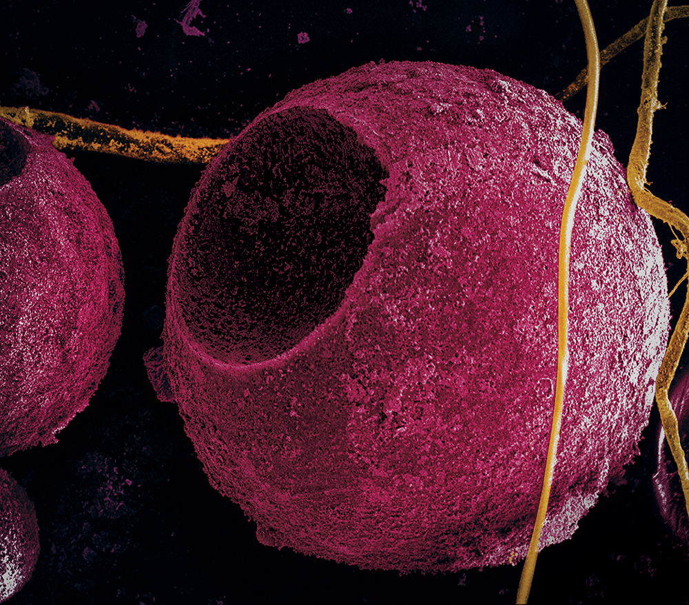
Quantum dots made of cadmium sulfide emit ultraviolet and blue wavelengths of light. Toxicity remains a concern for biological applications.
Featured in Asia Research News 2020 Magazine
The race is on to develop perfect quantum dots for looking inside cells and living tissues. These tiny, nanosized particles made from semiconductor materials fluoresce when exposed to light. This property makes them attractive for a variety of applications, including LED displays and solar cells. But for biological imaging, researchers still need to develop non-toxic quantum dots that can shine brightly deep inside tissues.
Quantum dots have been investigated for tagging tissues since 1998, but in vivo deep-tissue imaging is “still extremely limited, due to their toxicity to the environment and the human body,” says Naoto Shirahata, a chemist at Japan’s National Institute for Materials Science (NIMS).
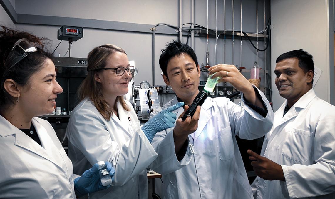
Shanmugavel Chinnathambi (right) and Naoto Shirahata (second from right) discuss with colleagues a key feature
needed to enhance the intensity of light emitted by their quantum dots.
Shirahata and Shanmugavel Chinnathambi, a research fellow at the NIMS International Center for Young Scientists, analysed the latest advancements in quantum dot biomarkers for the journal Science and Technology of Advanced Materials.
Near-infrared-emitting quantum dots made from cadmium, selenide, mercury, tellurium and lead exhibit bright luminescence, but their toxicity is a major drawback. Scientists have tried to overcome this problem by coating quantum dots made from these materials with a protective, non-toxic shell, yet the results were unsatisfactory.
Did you know?
Silicon quantum dots are a strong contender for safe bioimaging. They are non-toxic even at concentrations 15 times higher than leadbased quantum dots with just a silicon-oxide coating.
Silicon quantum dots, on the other hand, are non-toxic even at high concentrations, and have good tissue penetration under certain conditions. Scientists think they can improve this material by modifying its surface chemistry for highly selective tumour targeting. Germanium quantum dots are also interesting non-toxic candidates, but their emission is still too faint.
Shirahata, Shanmugavel and their colleagues are striving to improve the optical performance of non-toxic quantum dots. They are testing surface chemistry modifications to better target specific cells in the body. They also want to find ways to improve the body’s ability to safely metabolize quantum dots after their job is done.
“Silicon quantum dots in particular are promising,” says Shirahata, “because they are safe and can be metabolized with the urea cycle in the liver and the kidney.”
Further information
Naoto Shirahata | E-mail: [email protected]
International Center for Materials Nanoarchitectonics
National Institute for Materials Science
Shunichi Hishita | E-mail: [email protected]
Science and Technology of Advanced Materials
National Institute for Materials Science
Read this story in the Asia Research News 2020 magazine.
The many ways we can tell your research story. Find out more from our Content services page.
Advertisement