


Researchers at The University of Osaka have developed an improved technique called “tapping-mode scanning probe electrospray ionization” (t-SPESI), allowing the analysis of regions within individual cells with greater sensitivity and stability. Through device miniaturization and molecular modification of the probe surface, the team achieved higher sensitivity and long-term stability, successfully visualizing lipid distributions in mouse brain tissue at a pixel size of 5 µm.
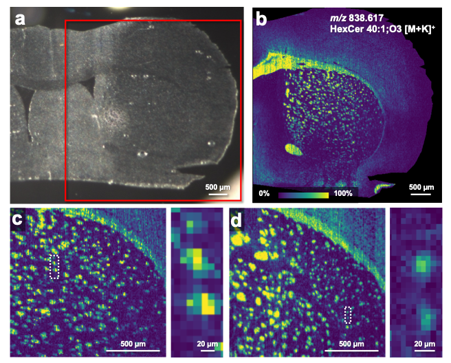
Fig. 3
(a) Optical microscope image of a mouse brain section. MSI was performed in the region indicated by the red box. (b) Ion image of m/z 838.617 acquired at a pixel size of 10 µm. (c) Enlarged ion image of the striatal region shown in (b). (d) Ion image of m/z 838.617 acquired at a pixel size of 5 µm.
©CC BY, Adapted from Figure 1 of "Development of a Tapping-Mode Scanning Probe Electrospray Ionization Platform for High-Sensitivity and Long-Term Stability in Single-Cell Mass Spectrometry Imaging of Tissue" by Yasuda et al., 2026, Analytical Chemistry, licensed under CC BY 4.0. DOI: 10.1021/acs.analchem.6c02386
Researchers from the University of Osaka have developed an improved analytical method that allows the interior of a tissue section to be examined at a single cell level with greater sensitivity and stability based on monitoring chemical components.
Osaka, Japan – Cells sitting side by side in the same tissues are not identical. Each cell carries its own subtly different chemical signature — a hidden individuality that can reveal how diseases take root and spread. Now, researchers from the University of Osaka have developed a technique sensitive enough to capture this cell-by-cell diversity within tissues, with unprecedented precision and stability.
Changes in the chemical makeup of cells can indicate the onset and progression of disorders such as neurodegenerative diseases, making it important to examine such changes in detail, focusing on the smallest possible areas. In the past, ambient sampling and ionization methods using electrospray ionization (ESI) for mass spectrometry imaging (MSI) has been developed.
ESI-based MSI uses a small probe to deliver solvent (a liquid that dissolves and releases chemical components) to a cell, detaching molecules that become charged and are then separated and counted in a mass spectrometer. Because mammalian cells can be as small as 10 micrometers, this imaging technique must be able to produce pixel sizes of less than this value.
“One issue with mass spectrometry imaging is that, as we focus on smaller and smaller regions within the cell, we require increasingly high sensitivity and stability,” says lead author Takao Yasuda.
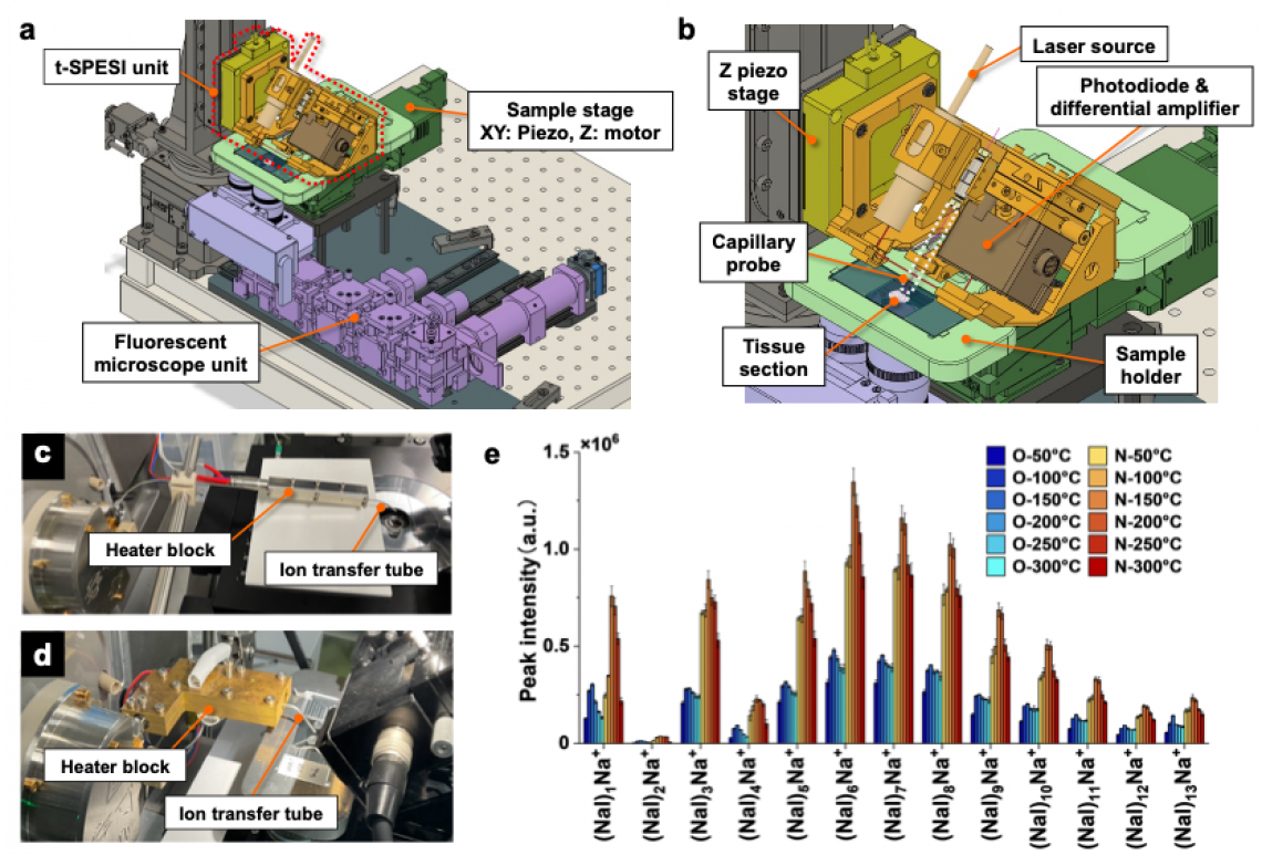
Fig. 1
Caption: (a) Rendered image of the developed measurement system. (b) Enlarged view of the t-SPESI unit and the sample stage unit. (c) Photograph of the conventional ion transfer tube. (d) Photograph of the developed ion transfer tube. (ed) Comparison of the signal intensities of NaI cluster ions. In the legend, O and N indicate the results obtained using the ion transfer tubes shown in (c) and (d), respectively, and the numbers indicate the heater temperature.
©CC BY, Adapted from Figure 1 of "Development of a Tapping-Mode Scanning Probe Electrospray Ionization Platform for High-Sensitivity and Long-Term Stability in Single-Cell Mass Spectrometry Imaging of Tissue" by Yasuda et al., 2026, Analytical Chemistry, licensed under CC BY 4.0. DOI: 10.1021/acs.analchem.6c02386
To address this issue, the researchers looked at ways to improve the performance of ESI-based MSI system named tapping-mode scanning probe ESI (t-SPESI), which was originally invented by a corresponding author, Yoichi Otsuka. In t-SPESI process, an extremely fine fused silica probe “taps” the cell repeatedly, alternately delivering a solvent and extracting components for analysis. This tapping motion enables the use of an extremely small amount of solvent to examine smaller areas but requires high sensitivity and good stability.
“Two factors currently limit the performance of this technique,” points out senior author Yoichi Otsuka. “These are the long pathway between the probe and the mass spectrometer, and the tendency for cell components to adhere to the probe surface over time.”
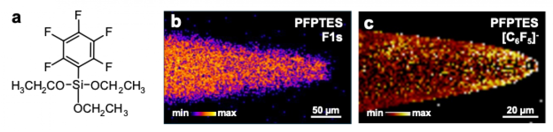
On this basis, higher sensitivity was realized by the research team through miniaturization of the complex analytical apparatus, reducing device mass by 45% and ion pathway length by 56%. Shortening the tube more than doubled the signal intensity. To ensure long-term stability by reducing the adhesion of sample to the probe, the silica probe surface was coated with a fluorine-containing chemical, somewhat like a nonstick coating on a kitchen implement.
As a test of this new system, mouse brain tissue samples were analyzed, and the team successfully visualized lipid distributions, including lipid classes previously implicated in Alzheimer’s and Parkinson’s disease, with a pixel size of 5 micrometers, corresponding to fine tissue structures, and with good stability.
The team expects that examining cells within tissues using this technology will provide new insights for disease research and treatment. With further optimization, e.g., of probe size, even better performance could be achieved, helping future studies uncover the mechanisms behind many disorders and advancing understanding of many disorders.
The article, “Development of a Tapping-Mode Scanning Probe Electrospray Ionization Platform for High-Sensitivity and Long-Term Stability in Single-Cell Mass Spectrometry Imaging of Tissue,” was published in Analytical Chemistry at https://doi.org/10.1021/acs.analchem.6c02386.
Fig. 2
(a) Chemical structure of PFPTES. (b) F1s intensity images obtained by XPS imaging of PFPTES-modified probes. (c) TOF-SIMS ion images of C6F5− obtained from PFPTES modified probes.
©CC BY, Adapted from Figure 1 of "Development of a Tapping-Mode Scanning Probe Electrospray Ionization Platform for High-Sensitivity and Long-Term Stability in Single-Cell Mass Spectrometry Imaging of Tissue" by Yasuda et al., 2026, Analytical Chemistry, licensed under CC BY 4.0. DOI: 10.1021/acs.analchem.6c02386
About The University of Osaka
The University of Osaka was founded in 1931 as one of the seven imperial universities of Japan and is now one of Japan's leading comprehensive universities with a broad disciplinary spectrum. This strength is coupled with a singular drive for innovation that extends throughout the scientific process, from fundamental research to the creation of applied technology with positive economic impacts. Its commitment to innovation has been recognized in Japan and around the world. Now, The University of Osaka is leveraging its role as a Designated National University Corporation selected by the Ministry of Education, Culture, Sports, Science and Technology to contribute to innovation for human welfare, sustainable development of society, and social transformation.
Website: https://resou.osaka-u.ac.jp/en
Advertisement