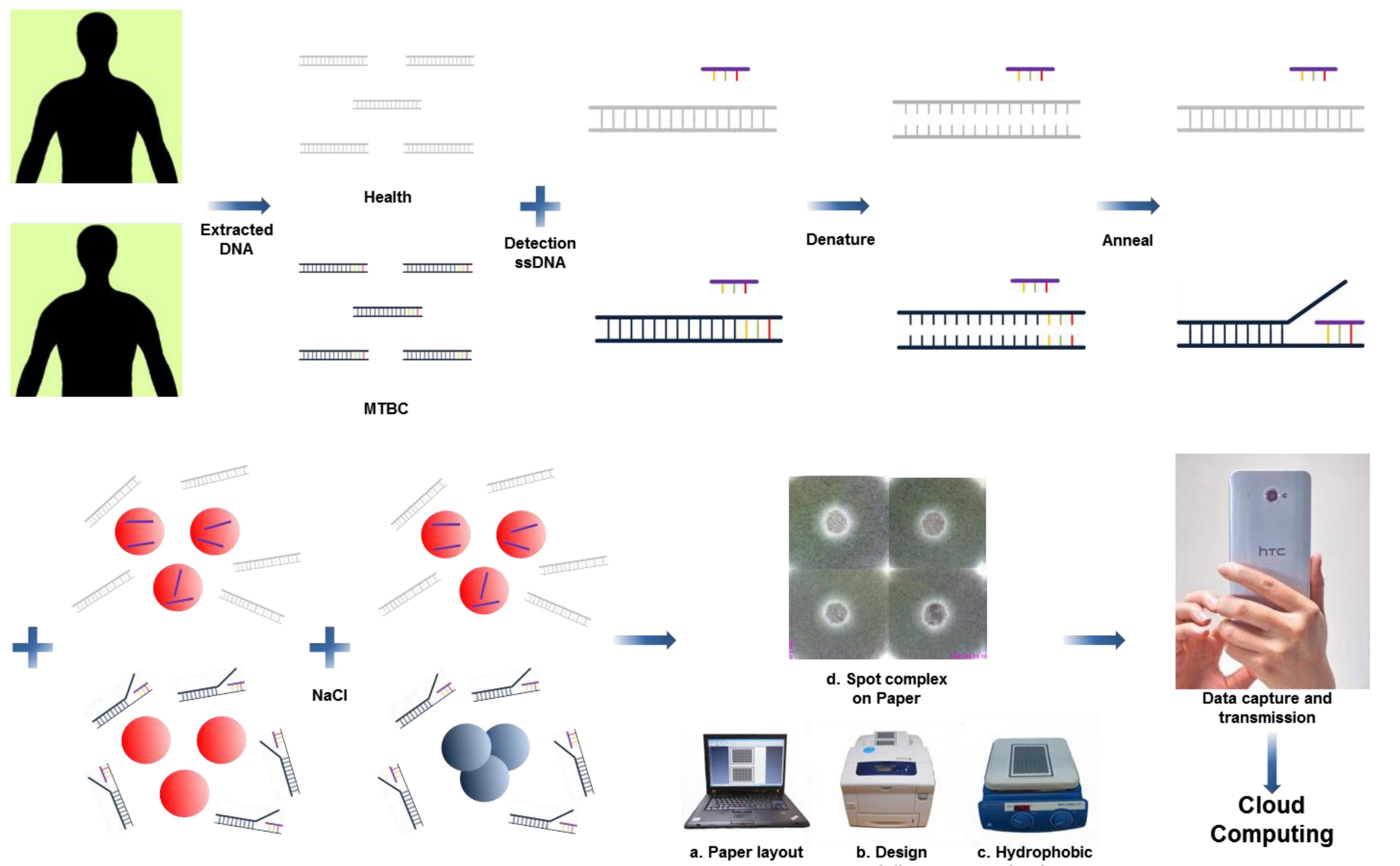
Figure 1. Schematic of the proposed TB diagnosis. Unknown extracted human DNA sequences are first hybridized with detection oligonucleotide sequences, followed by addition of colloidal gold nanoparticles and triggering of the colorimetric sensing with a sodium chloride solution. If the extracted DNA sequences consist of IS6110 target sequences then the detection oligonucleotide sequences will hybridize with them, and only a few ssDNA sequences will be absorbed on the gold nanoparticles to avoid aggregation after the addition of salt. In the absence of IS6110 target sequences, the color of the mixture remains red after hybridization and does not change. The mixture is then spotted and concentrated on the chromatography paper confined by solid wax hydrophobic barriers. Diagnostic results can be photographed with a smartphone and sent to a server for cloud computing.
More than a century after the identification of organisms that cause tuberculosis (TB), this disease remains a global public health challenge. According to World Health Organization estimates, there were 8.7 million new cases in 2011 and 1.4 million deaths. Most new cases occur in developing countries that lack the facilities and trained personnel required for early detection of TB.
In a new study, published in the journal Science and Technology of Advanced Materials (STAM), researchers in Taiwan describe a simple, color-based diagnostic approach with the potential to detect target DNA sequences found in TB-causing mycobacteria – in just a fraction of the time required for established diagnostic tests.
The standard method for TB detection in a clinical setting involves culturing the Mycobacterium tuberculosis bacillus, which requires 3-6 weeks to grow on solid culture media or 9-16 days in rapid liquid culture media. A faster alternative is the polymerase chain reaction (PCR) technology. However, it is still too slow (turnaround time 2-5 hours) and requires sophisticated infrastructure and trained personnel that might be unavailable in developing countries.
In their STAM paper, Tsung-Ting Tsai and colleagues employed gold nanoparticles and microfluidic paper-based analytical devices to achieve rapid diagnosis without the need for complex and time-consuming laboratory processes. They easily detected TB mycobacterium target sequences, and the turnaround time was approximately 1 hour after the human DNA was extracted from patients.
Although the authors are still optimizing their technology, they already believe that it will result in “affordable, sensitive, specific, user-friendly, rapid and robust, equipment-free, and highly end-user-deliverable diagnostic applications”.
For more information about this research, please contact:
Media contacts:
National Institute for Materials Science, Tsukuba, Japan
Email: [email protected]
Tel. +81-(0)29-859-2494