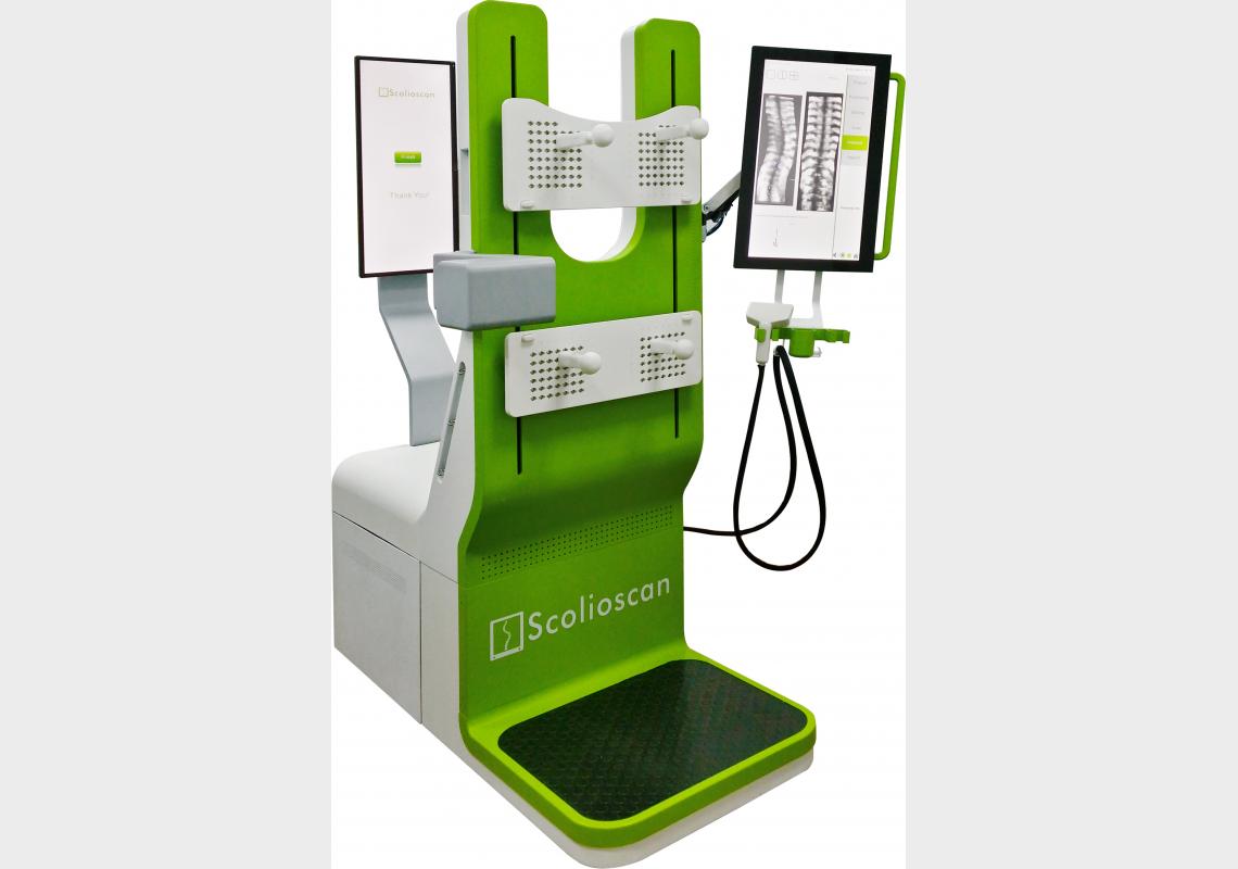
Unlike X-rays, the Scolioscan is radiation-free and enables safer screening whenever needed.
Scoliosis is a medical condition that is defined as a three-dimensional spine deformity with curvature of more than ten degrees in the coronal plane (a vertical plane that divides the body into its front and back halves). For young teenagers, the condition – known as adolescent idiopathic scoliosis (AIS) – can progress to a large curvature that is six times greater than that in people above 16 years of age, as their bodies are still in the process of developing and hence are more vulnerable to curvature.
Although regular screening can help prevent scoliosis from worsening, the most common diagnostic method uses Xrays. Due to the health risks posed by radiation exposure – including an increased incidence of lung and breast cancers – regular screening cannot be conducted too frequently, particularly in young patients. Previous studies indicate that X-ray images for AIS patients may be taken every 3-12 months for 3-5 years.
Led by Professor Yongping Zheng, researchers at The Hong Kong Polytechnic University have developed “Scolioscan”, a radiation-free technology that can accurately assess scoliosis in three dimensions.
The Scolioscan can assess a patient’s spine in the standing posture and generate coronal images of spinal curvature, which can then be measured to determine the severity of the scoliosis. The technology captures the spine’s 3D profile using identified bony landmarks to measure spinal rotation and deformity along different planes.
Unlike X-rays, the Scolioscan is radiation-free and enables safer screening whenever needed, allowing healthcare workers to detect scoliosis at an early stage or avoid unnecessary treatments for patients with stable spinal angles. It also allows close follow-up monitoring on the progress of spinal bracing or other treatments. “This technology can reduce the economic and psychological burdens for patients and their families,” says Professor Zheng.
Professor Zheng and his colleagues are currently exploring the Scolioscan’s potential. The team aims to examine the spine’s structural development during childhood and establish baseline data on the prevalence of scoliosis at different age groups, while improving the technology’s functionality. For example, they hope to make the measurement process quicker, more automatic and more accurate as well as add angle measurements along the sagittal plane (a vertical plane that divides the body into its right and left halves).
Another goal is to verify the results produced by Scolioscan assessments. “The current verification standard is based on the Cobb’s angle method [used to measure spinal deformity] but it does not always represent the true value of spinal curvature,” notes Professor Zheng.
“The values of spinal curvature produced by Cobb’s angle can vary by up to seven degrees. Therefore, further research is needed to find a better way to verify the angles measured by the Scolioscan, or ultimately establish its own standard,” he concludes.
For further information contact:
Professor Yongping Zheng
Interdisciplinary Division of Biomedical Engineering
The Hong Kong Polytechnic University
E-mail: [email protected]
*This article also appears in Asia Research News 2015 (P.38).


