

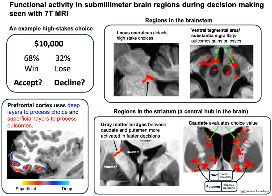

Scientists have identified submillimeter gray matter nuclei in the human brain involved in the distinct stages of processing information when making rapid, high-stakes decisions. These findings provide critical insights for precise targeting of the neural substrates that govern human choice behavior.
Mesoscale neuroimaging of human decision processing.
National Taiwan University, Joshua Oon Soo Goh; University of Illinois Urbana-Champaign, Yuhui Chai, Bradley Sutton.
Imagine that a notification on your financial app captures your attention about a stock projected to have a 68% chance of doubling your investment within the month, with a 32% chance of total loss. Would you commit a $50,000 stake? Would your decision change if the odds of gain rose to 99 to 1, or if the stake dropped to $1,000? Even after your decision, you would likely track the stock outcome to see if you can fund that family vacation, incur debt, or merely maintain your financial status.
The above scenario depicts a value-based choice to accept or avoid risks. Such choices permeate everyday life from routine tasks to high-stakes decisions affecting well-being and survival. Because the outcomes of these choices are inherently uncertain, the brain integrates multistage neural information processing in order to select adaptive actions and prevent potential harm. A precise understanding of these neural circuits is essential for addressing maladaptive decision behaviors in neuropsychiatric or neurodegenerative conditions.
In a collaborative study, researchers at National Taiwan University (NTU) and the University of Illinois at Urbana-Champaign (UIUC) have identified submillimeter gray matter nuclei in the human brain involved in the distinct stages of processing information when making rapid, high-stakes decisions. Published in Nature Communications, these findings offer critical insights for precise targeting of the neural substrates that govern human choice behavior.
The researchers scanned participants using a 7-Tesla (7T) ultra-high-field magnetic resonance imaging (MRI) system while they performed a specialized decision-making task. The ultra-high field strength allowed the team to overcome the spatial resolution limitations of conventional 3-Tesla (3T) scanners, providing an unprecedented look at the functional activity of mesoscale brain structures – ranging from hundreds of microns to one millimeter in size – during human decision-making.
“We previously developed the lottery decision task for 3T systems, which resolve neural activity at roughly 3 mm resolution to reveal broad brain areas generally sensitive to different stake values or decision certainty,” says co-corresponding author Dr. Joshua Goh, associate professor at Graduate Institute of Brain and Mind Sciences, National Taiwan University.
“By tuning our task stimuli to specifically target mesoscale structures at 7T, we could disentangle a series of small structural hubs in the brain that act as functional gates for different kinds of information during a decision. At 3T resolution and with less optimized tasks, these fine-grained functional distinctions would likely remain hidden.”
In the study, the researchers found that when salient high stakes were encountered, the locus coeruleus – a slender brainstem structure about 2 mm across – signaled the rest of the brain, particularly the frontal cortex, to allocate heightened neural resources for processing the critical decision rather than continuing baseline operating levels. Within the 3 mm-thick frontal cortex, neural engagement shifted between the deeper layers during choice formulation and the superficial layers during outcome evaluation.
To further process outcomes, the ventral tegmental area/substantia nigra (VTA/SN) in the brainstem and nucleus accumbens (NAcc) in the striatum – both millimetric structures important in dopamine reward signaling – differentiated gains from losses, reflecting their role in updating future behaviors.
Crucially, the researchers also identified a novel functional role for gray matter bridges (GMBs) – structures about 1 mm wide spanning the striatum between the caudate and putamen. GMB activity was higher for faster decisions than slower ones. This pattern suggests that GMBs act as an information gate: signaling when accumulated evidence is sufficient to pass downstream for action execution in the putamen, or whether extended upstream processing is required back in the caudate or cortex.
“A key MRI methodological advancement was developing a technique to acquire functional and structural brain images simultaneously, rather than in separate scans as is conventional,” says first and co-corresponding author Dr. Yuhui Chai, a research scientist of Beckman Institute for Advanced Science and Technology at University of Illinois Urbana-Champaign. “This is critical for submillimeter precision, as even the slightest misalignment that might occur between scans can drastically compromise the precise spatial localization of functional activity in these mesoscale structures.”
When brain functions break down – such as through age-related neurodegeneration or neurological pathology – the deterioration likely originates in discrete neural microcircuits rather than across whole brain regions. Mapping and quantifying how these mesoscale decision circuits operate in living humans is a major milestone. It allows us to detect individual vulnerabilities earlier and develop more targeted interventions before impaired decisions lead to severe life-altering consequences.
“This allows us to map the brain’s internal ‘assembly line’ – from the moment valuable information is detected, through evidence integration to reach a decision, to execution of an action, and the evaluation of the outcomes,” adds Dr. Joshua Goh. “With this detailed framework, we have greater confidence in pinpointing which part of the assembly line is disrupted when human decision-making begins to falter.”
Prof. Joshua Oon Soo Goh's email address: [email protected]
The Mind and Brain Laboratory of Prof. Goh linked to https://gibms.mc.ntu.edu.tw/bmlab/people/joshua-goh.html
Advertisement