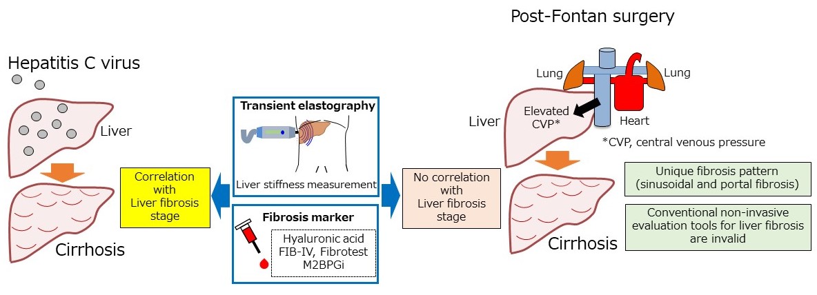
Conventional non-invasive evaluation tools for liver fibrosis post-Fontan surgery are invalid
It is well-known that patients who undergo Fontan surgery slowly develop liver fibrosis for years post-operatively. In the past decade, these incidences have been steadily increasing and this is due partly to the need for an accurate diagnostic method. A research group led by Dr. Daisuke Tokuhara, Associate Professor of Pediatrics, Osaka City University Graduate School of Medicine and Dr. Yuki Cho have found that the conventional methods of ultrasound elastography and biomarkers via blood tests do not show the actual status of postoperative liver fibrosis. Their findings have been published in Hepatology Research.
With a sample size of 22 postoperative patients, their illumination into this matter was 3-prong.
Frist, the team observed dilation of the sinusoid during a long-term period post-Fontan surgery, with moderate fibrosis extending from the sinusoidal region to the portal vein region in about half of the patients, and a severe form developing in 30% of the patients. “After Fontan surgery, we believe as central venous pressure increases and the sinusoids dilate, fibrosis spreads from the sinusoidal region to the portal vein region”, states Professor Tokuhara. Contrast this with hepatitis C and B, which are well-known causes of cirrhosis, where fibrosis is observed mainly in the portal region.
Then the team tested this unique regional development of fibrosis. Using a method of investigation that measures liver stiffness called ultrasound elastography, they found that the score did not significantly relate to liver fibrosis after Fontan surgery because the liver hardness tended to be related to portal pressure.
Next, they diagnosed liver fibrosis by looking for the elements that are naturally present as a result of the diseases progression. The research team investigated a variety of these fibrosis biomarkers from blood tests. Analysis of hyaluronic acid, type 4 collagen 7s, APRI, FIB-4 Index, and FibroTest did not render any association with the liver fibrosis score after Fontan surgery. The biomarker M2BPGi showed a significant inverse U-shaped association with postoperative liver fibrosis score, “but this cannot be used in a clinical practice”, says Dr. Cho.
The team concluded that the complication with long-term care after Fontan surgery lies in a specific liver fibrosis that is difficult to assess using conventional methods. “Postoperative patients should receive regular medical care at specialized medical institutions for the presence of liver complications for at least 10 years after surgery, and until useful biomarkers and imaging techniques are established”, advises Dr. Tokuhara. “We will use the liver tissue and serum obtained from this study to continue our search for the world`s first biomarker for liver fibrosis specific to post-Fontan surgery.”
###
We are Osaka City University - the oldest research university in Osaka. With 9 undergraduate faculties and 11 graduate schools all dedicated to making urban life better, energy cleaner, and people healthier and happier, we have won numerous awards and have produced 2 Nobel laureates. For more information, please visit our website at https://www.osaka-cu.ac.jp/en