

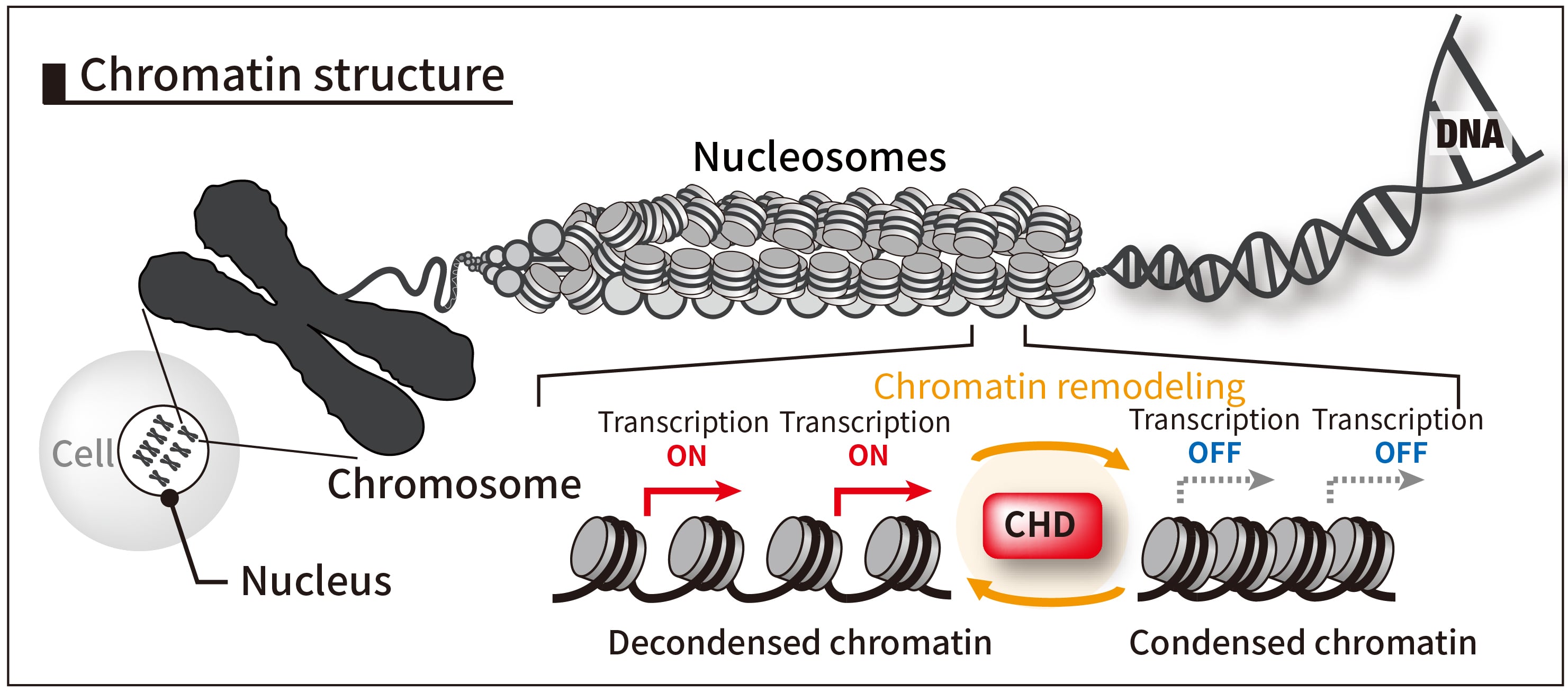
Figure 1. CHD8-induced chromatin remodeling.
In chromatin found in the nucleus, DNA is highly folded and packed into structures known as nucleosomes, where the DNA is wrapped around proteins called histones. Gene expression is regulated by the decondensation and condensation of the chromatin. CHD8 possesses chromatin remodeling activity that alters chromatin structure, thereby regulating the switching of gene transcription between on and off states.
Background
Autism spectrum disorder (hereafter, autism) is a developmental disorder characterized by difficulties with social-interaction and by restricted interests and repetitive behaviors. It has attracted significant attention due to its high prevalence, affecting approximately one in every 36 persons, and symptoms that interfere with daily life. In recent years, genetic mutation analysis targeting individuals with autism has revealed that CHD8*1), a chromatin remodeler, is one of the most frequently mutated genes associated with autism, attracting significant attention (Figure 1).
CHD8 is known to be involved in the differentiation and maturation of diverse cells in the brain. It was unknown, however, at which stage of brain development and in which types of cells mutations in the CHD8 gene exert their effects to cause autistic-like behavior.
Results
This study was conducted by a team of researchers from the Institute of Frontier Science Initiative and from the Institute of Medical, Pharmaceutical and Health Sciences, Kanazawa University. First, they succeeded in generating mice in which the expression level of CHD8 could be reduced at a specific developmental stage, using gene-editing technology. As a result, it was revealed that mice with reduced CHD8 expression at the midfetal stage (embryonic day 14.5) exhibited autistic-like behavior, such as abnormal social-interaction and anxiety-like behavior, while such behavioral abnormalities were not observed in mice with reduced CHD8 expression at a late fetal stage (embryonic day 17.5) onwards (Figure 2). Furthermore, through gene expression analysis and histological analysis of the brain, they determined that CHD8 mutation at the midfetal stage excessively promoted the differentiation of ventral progenitor cells*2), thereby causing developmental abnormalities in inhibitory neurons*3) and oligodendrocyte*4)-lineage cells. In addition, using spatial transcriptomics and in vivo neural circuit functional analysis*5), they revealed that changes in inhibitory neurons associated with CHD8 mutation led to functional abnormalities in neural circuits in the adult brain (Figure 3).
Moreover, they demonstrated that genetic restoration of CHD8 expression at a fetal stage ameliorated both the abnormal differentiation of ventral progenitor cells and the behavioral abnormalities in mice.
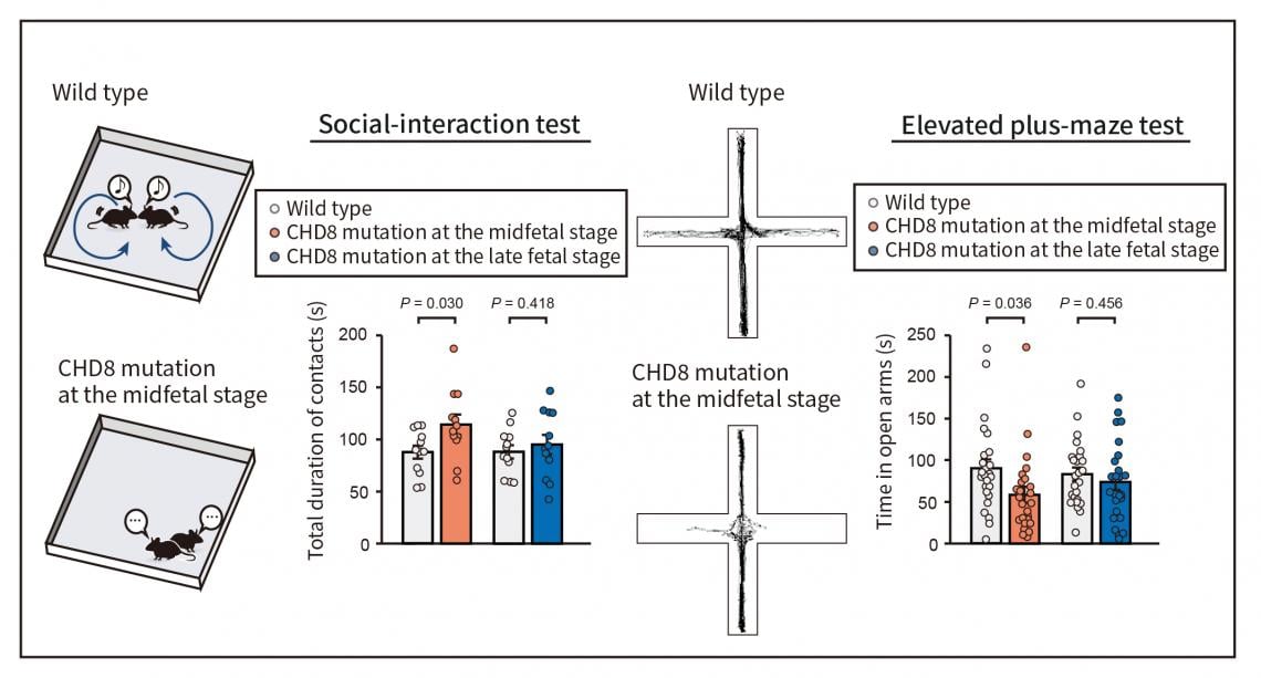
Figure 2. Behavioral abnormalities due to a CHD8 mutation at the midfetal stage.
Mice were generated that develop a CHD8 mutation at a specific fetal stage and behavioral analysis was conducted after birth. As a result, it was revealed that mice with a CHD8 mutation induced at the midfetal stage (embryonic day 14.5) exhibited autistic-like behavior (abnormal social-interaction, left panel; anxiety-like behavior, right panel), while mice with a CHD8 mutation induced at the late fetal stage did not exhibit behavioral abnormalities.
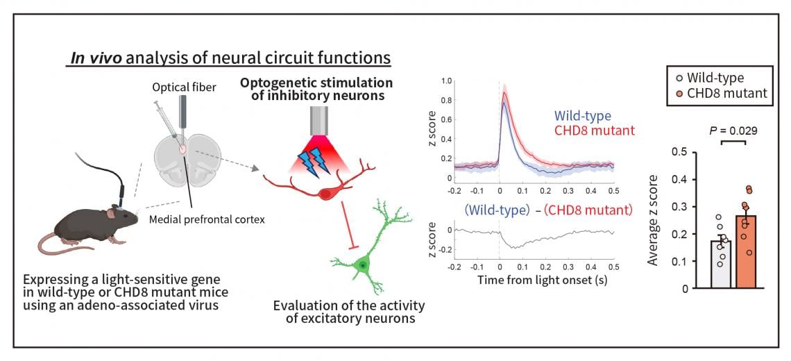
Figure 3. Abnormalities in inhibitory neural circuits arise in association with the CHD8 mutation.
The team analyzed in vivo neural circuit function by expressing a light-sensitive gene in inhibitory neurons of the mouse medial prefrontal cortex and applying optical stimulation to that region (left panel). As a result, it was revealed that in CHD8-mutant mice, excitatory neurons were less susceptible to inhibition even when inhibitory neurons were stimulated (right panel).
Future prospects
While CHD8 has attracted attention as a major autism-associated gene, it remained unclear when, where, and in which cell types it contributes to the behavioral abnormalities underlying the core symptoms of autism. The present study revealed that the neurodevelopmental differences at the root of the behavioral abnormalities stem from aberrant differentiation of ventral progenitor cells at the midfetal stage (Figure 4). The present findings are expected to significantly advance our understanding of the mechanism underlying the onset of autism and pave the way for the development of new therapeutic strategies targeting specific neurodevelopmental stages or cell types.
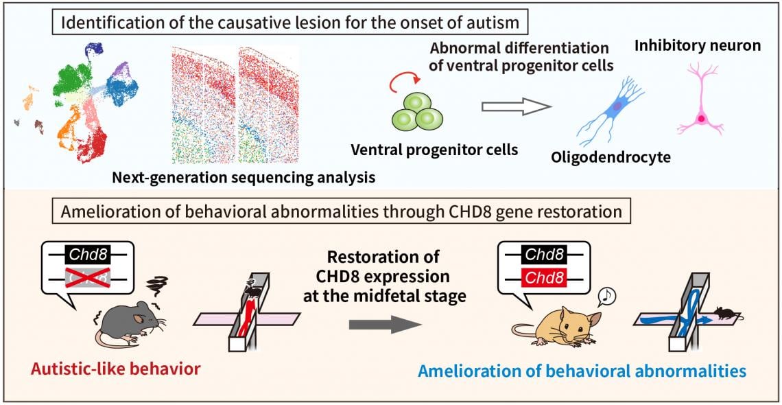
Figure 4. Elucidation of the mechanism underlying autism onset through the CHD8 mutation and therapeutic applications.
Next-generation sequencing analysis on cells harboring the CHD8 mutation at the midfetal stage revealed that abnormalities occurring when ventral progenitor cells differentiated into oligodendrocytes and inhibitory neurons were closely related to the onset of autistic-like behavior (upper panel). In addition, when CHD8 expression levels were restored at the midfetal stage in autism model mice, behavioral abnormalities were ameliorated (lower panel). These findings are expected to contribute to the elucidation of the pathophysiology of autism and to applications in its treatment.
Glossary
*1) CHD8
Abbreviation of Chromodomain Helicase DNA binding protein 8, a protein belonging to a group known as chromatin remodelers, which utilize intracellular energy to alter chromosome structure and regulate gene expression levels.
*2) Ventral progenitor cells
Ventral progenitor cells are undifferentiated cells that can differentiate into neurons or glial cells during brain development. In particular, neural progenitor cells located in the ventral region of the brain serve as the origin of cells, such as inhibitory neurons and oligodendrocytes, which are crucial for regulating the function of neural circuits.
*3) Inhibitory neuron
Inhibitory neurons are those within neural circuits that suppress the activity of other neurons. They primarily use the neurotransmitter GABA to suppress excessive neuronal excitation and maintain a balance of information processing within the brain.
*4) Oligodendrocyte
Oligodendrocytes are a type of glial cell in the brain and spinal cord that form the myelin sheath covering the axons of neurons. The myelin sheath increases the conduction velocity of nerve signals and is essential for the proper functioning of neural circuits.
*5) In vivo analysis of neural circuit functions
An analytical method for investigating how neural circuits function by stimulating and recording from specific neurons or neural circuits in the brain of living animals. In this study, the presence or absence of abnormalities in neural circuits was evaluated by selectively stimulating specific neurons and observing the effects.
###
About Kanazawa University
— Contributing to Society through “Future Oriented Intelligence”, Built on an “All Kanazawa University” Approach.
Kanazawa University (KU), founded in 1862, is a research university dedicated to education, while opening up its doors to both local and global society. Guided by our vision, “Kokorozashi,” we contribute to society through “Future-oriented Intelligence,” addressing current challenges and anticipating future ones from both local and global perspectives.
KU includes 4 colleges, 20 schools, 7 graduate schools, a hospital, and specialized research centers such as the Cancer Research Institute, a leading hub for research on cancer metastasis and drug development. Over 1,000 researchers drive innovation and international collaboration across diverse fields.
KU is advancing research through WPI (World Premier International Research Center Initiative) and J-PEAKS (Program for Forming Japan’s Peak Research Universities), accelerating interdisciplinary and international collaborations and innovations.
Learn more here: https://www.kanazawa-u.ac.jp/en/