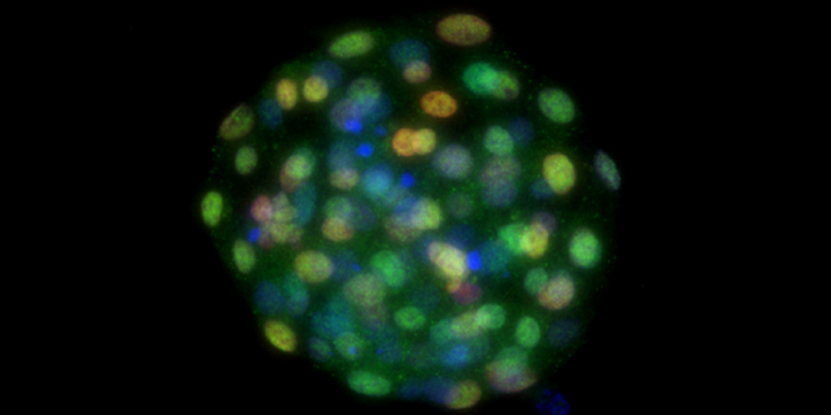
Mammalian development starts from a single cell — a fertilized egg. The egg goes through multiple cell divisions to increase its cell numbers and then starts forming a sphere-like structure with a cavity inside, called the blastocyst. The blastocyst consists of two types of cells, the inner cell mass (ICM) and the trophectoderm (TE), which develop into an embryo proper and a large part of the placenta, respectively.
Scientists led by Manabu Kawahara at Hokkaido University have shown that, since bovine ICM cells can regenerate TE, they are capable of forming both the embryo and placenta. The study was published in the Journal of Biological Chemistry and became one of the top 50 most viewed papers from November through December 2019 on the Journal’s website.
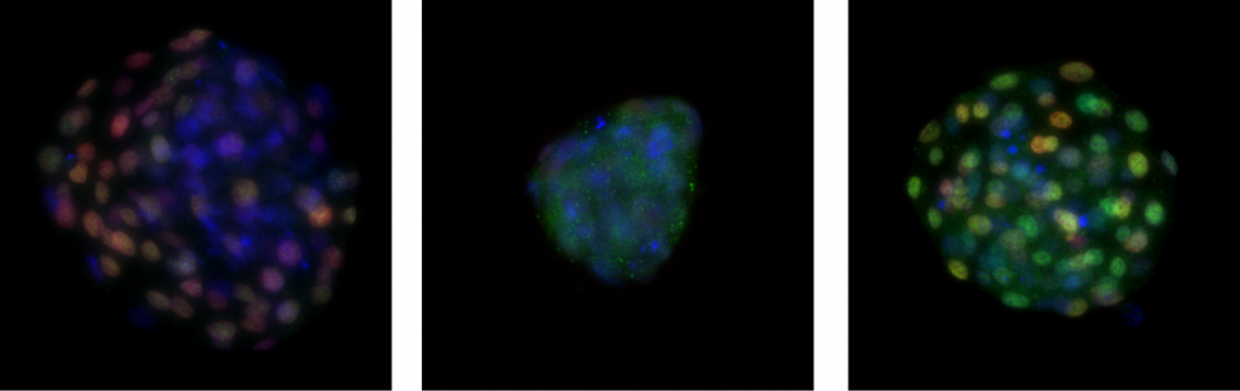
An early bovine embryo regenerating its TE cells which will later form a large part of the placenta. (Left: intact, Middle: after removal of TE, Right: regenerated) (Kohri N. et al., Journal of Biological Chemistry. November 8, 2019)
To examine the ICM’s capacity to regenerate TE, the researchers cultivated mouse and bovine blastocysts and removed entire TE from both blastocysts. They found that both blastocysts regained their sphere-like shapes in 24 hours. However, the regeneration rate to reform the blastocyst was remarkably higher in bovine cells (97%) than mouse cells (57%). The more complete recovery of bovine blastocysts in cell numbers compared to mouse blastocysts suggests the bovine cells have a higher regenerative capacity.
Further experiments revealed abnormal protein expression in the TE of mouse regenerated blastocysts, whereas bovine regenerated blastocysts showed normal gene expressions overall.
To test its developmental abilities, the researchers then transferred the regenerated blastocysts to recipient females. After the embryo-transfer, to their surprise, one of the four cows became pregnant and a female calf was naturally born with an apparently normal placenta. In contrast, none of the more than 100 mouse embryos transferred to recipients developed to term.
“We will continue to monitor the health of the calf born from the regenerated blastocyst,” says Manabu Kawahara. “Our study suggests that we can remove and use a large part of TE for genetic testing to breed cattle with improved qualities. Also, further studies could reveal the mechanism of cell fate decision in mammals and its differences between species.”
Contacts:
Associate Professor Manabu Kawahara
Laboratory of Animal Breeding and Reproduction
Research Faculty of Agriculture
Hokkaido University
Email: k-hara[at]anim.agr.hokudai.ac.jp
Naoki Namba (Media Officer)
Institute for International Collaboration
Yuki Otani (Science Writing Intern)
Public Relations Division
Hokkaido University
Tel: +81-11-706-2185
Email: en-press[at]general.hokudai.ac.jp
Paper: https://www.jbc.org/content/early/2019/11/08/jbc.RA119.010746