Graphical abstract
Cells have surface receptors that couple to proteins and other molecules to initiate or inhibit certain behaviors. Typically, the number of these receptors increases as the cell matures, but researchers have now identified that one receptor influences cell behavior much earlier than previously thought and appears to help trigger the cell differentiation process to form neurons.
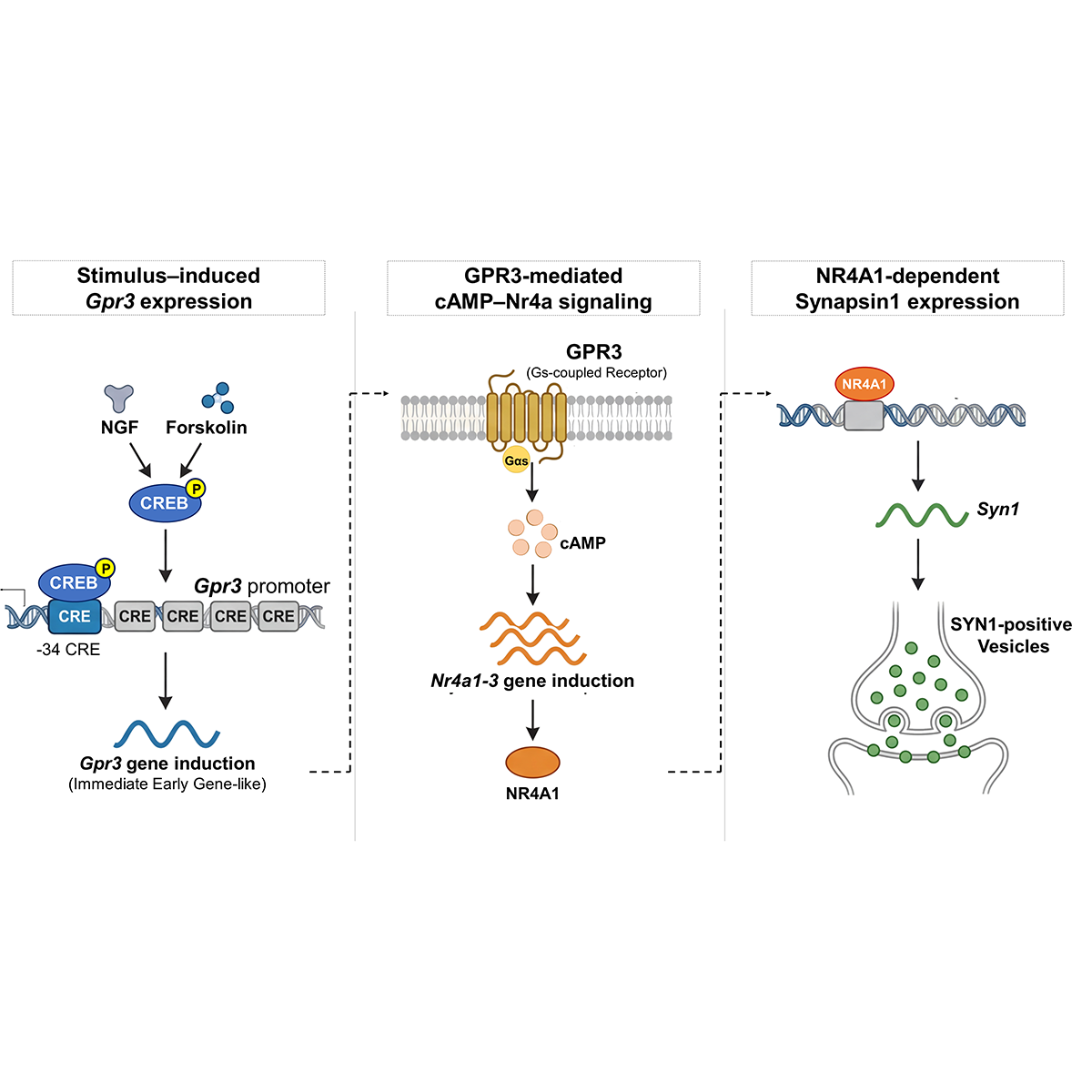
The Hiroshima University-based team published their work, which they said has implications for better understanding neuronal development and brain plasticity — and how those processes become dysregulated — on March 20 in iScience. They specifically found that G protein-coupled receptor 3 (GPR3) represents a unique molecule in this receptor family, as it behaves like an immediate-early gene that rapidly responds and induces downstream signaling. Other G protein-coupled receptors behave like delayed-response genes that aren’t expressed into much later in the cell maturation process.
“Understanding early transcriptional responses — how genes are expressed in response to upstream signals — is critical because these programs determine neuronal development, synaptic formation and plasticity, and their dysregulation is associated with neurological disorders such as autism and cognitive dysfunction,” said corresponding author Shigeru Tanaka, associate professor of molecular and pharmacological neuroscience in the Graduate School of Biomedical and Health Sciences at Hiroshima University.
Like a baseball player raising a gloved hand to catch a baseball, cell surface receptors extend out, waiting to receive specific molecules. When the baseball hits the mitt, it can trigger a series of reactions, depending on where the baseball originated. It can immediately end the play, or the catcher can use it to tap out an opponent, or throw it to a teammate who might be closer to the player who hit it in the first place. Just like the game can end or continue depending on how and where the ball moves, so can cell differentiation and behavior. However, many rules of play for cells in development still remain unclear, according to Tanaka. Making the situation even more complex is that GPR3 — the mitt — can exert its function even without a baseball, or a molecule to trigger a specific action.
To better understand how GPR3 works in the process, Tanaka and the research team analyzed rodent PC12 cells, a widely used and well-established scientific model for studying how cells differentiate into neurons. This neuronal differentiation process involves stimulating the cells with nerve growth factor, a signal that tells the cells to become neurons. Over 48 hours, the cells develop neurites, which are immature branches that may eventually form neuron networks if properly supported. The team then inspected the neuronal protein markers on the cells and found that GPR3 activated within 30 minutes of stimulation.
“That was striking — that GPR3 is one of the very few G protein-coupled receptors showing immediate-early gene-like rapid induction within 30 minutes,” Tanaka said. “That’s comparable to classical immediate-early genes, yet unprecedented for this receptor family.”
Tanaka explained that GPR3 could be considered a “signal amplifier,” meaning it converts early stimuli signaling from other molecules upstream into a sustained program necessary for neuronal maturation. Because GPR3 can act on its own, its early appearance may be especially important in helping this process happen quickly. Specifically, he said, genetic analysis revealed early induction of GPR3 enhances cAMP-CREB signaling, which makes long-term processes from short-term signaling. That, in turn, drives downstream expression of NR4A, an immediate-early gene critical for neuronal survival and the development of synapses, which are the spaces over which neurons communicate.
“This work establishes a previously unrecognized signaling cascade linking early transcriptional responses to synapse development,” Tanaka said.
Next, the researchers said they plan to investigate how GPR3 contributes to synaptic function, neural circuit formation and neurological disorders.
“Our ultimate goal is to clarify how activity-dependent transcriptional programs regulate brain development and to identify new therapeutic targets for neurodevelopmental and neuropsychiatric diseases,” Tanaka said.
Other contributors to the study are Fumiaki Ikawa, Hiroko Shiraki, Kana Harada, Izumi Hide and Norio Sakai, all of whom are affiliated with the Department of Molecular and Pharmacological Neuroscience in Hiroshima University’s Graduate School of Biomedical and Health Sciences.
The Japan Society for the Promotion of Science supported this research.
###
About Hiroshima University
Since its foundation in 1949, Hiroshima University has striven to become one of the most prominent and comprehensive universities in Japan for the promotion and development of scholarship and education. Consisting of 12 schools for undergraduate level and 5 graduate schools, ranging from natural sciences to humanities and social sciences, the university has grown into one of the most distinguished comprehensive research universities in Japan. English website: https://www.hiroshima-u.ac.jp/en