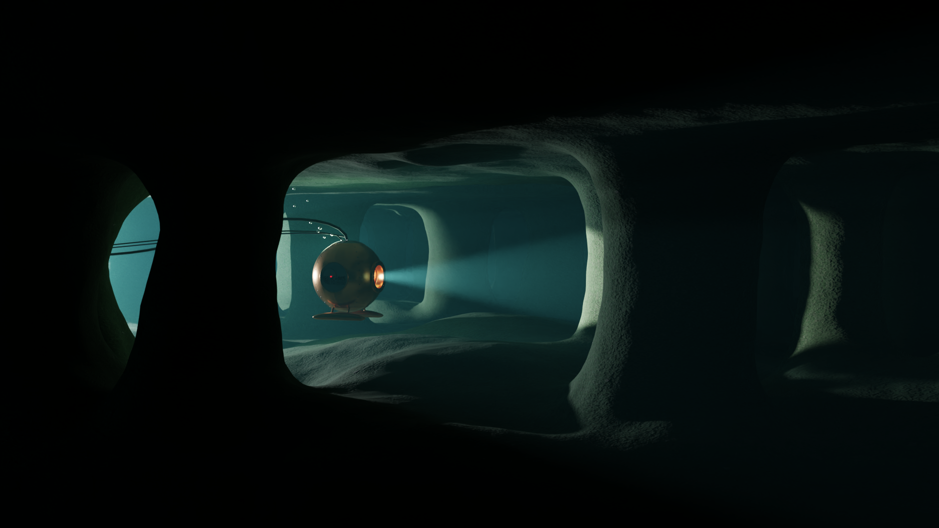
Exploration of a new structure for microfluidic devices. The image is an artistic rendering of the internal porous structure that is made using polymers and light. Researchers at iCeMS have achieved controlled liquid flow in this structure.
Microfluidic devices use tiny spaces to manipulate very small quantities of liquids and gasses by taking advantage of the properties they exhibit at the microscale. They have demonstrated usefulness in applications from inkjet printing to chemical analysis and have great potential in personal medicine, where they can miniaturize many tests that now require a full lab, lending them the name lab-on-a-chip.
Researchers at Kyoto University’s Institute for Integrated Cell-Material Sciences (iCeMS) approached microfluidic fabrication from a new direction and came up with an innovative process to make devices with some distinctive properties and advantages.
A description of the new process created by Dr. Detao Qin of iCeMS’ Pureosity team, led by Professor Easan Sivaniah, appears in Nature Communications.
Until now, making devices with microfluidic channels has required assembling them from several components, introducing possible points of failure. The Pureosity team’s process needs no such assembly. Instead, it uses light-sensitized common polymers and micro-LED light sources to create self-enclosed, porous, high-resolution channels, capable of carrying aqueous solutions and separating small biomolecules from each other, through a novel photolithography technique.
The Pureosity team’s latest development builds upon their Organized Microfibrillation (OM) technology, a printing process which was previously published in Nature (2019). Due to a unique feature of the OM process the microfluidic channels display structural color which is linked to the pore size. This correlation ties flow rate to the color as well.
“We see great potential in this new process,” says Prof. Sivaniah. “We see it as a completely new platform for microfluidic technology, not just for personal diagnostics, but also for miniaturized sensors and detectors.”
Microfluidic devices are already being used in the biomedical field in point-of-care diagnostics to analyze DNA and proteins. In the future, devices may allow patients to monitor their vital health markers by simply wearing a small patch, so that healthcare providers can respond immediately to dangerous symptoms.
“It was exciting to finally use our technology for biomedical applications,” says Assistant Professor Masateru Ito, a co-author on the current paper. “We are taking the first steps, but it is encouraging that relevant biomolecules such as insulin and the SARS-COV2 shell protein were compatible with our channels. I think that diagnostic devices are a promising future for this technology.”
###
DOI:
https://doi.org/10.1038/s41467-022-29956-4
About Kyoto University’s Institute for Integrated Cell-Material Sciences (iCeMS):
At iCeMS, our mission is to explore the secrets of life by creating compounds to control cells, and further down the road to create life-inspired materials.
https://www.icems.kyoto-u.ac.jp/
For more information, contact:
Christopher Monahan / I. Mindy Takamiya
[email protected]
Advertisement