

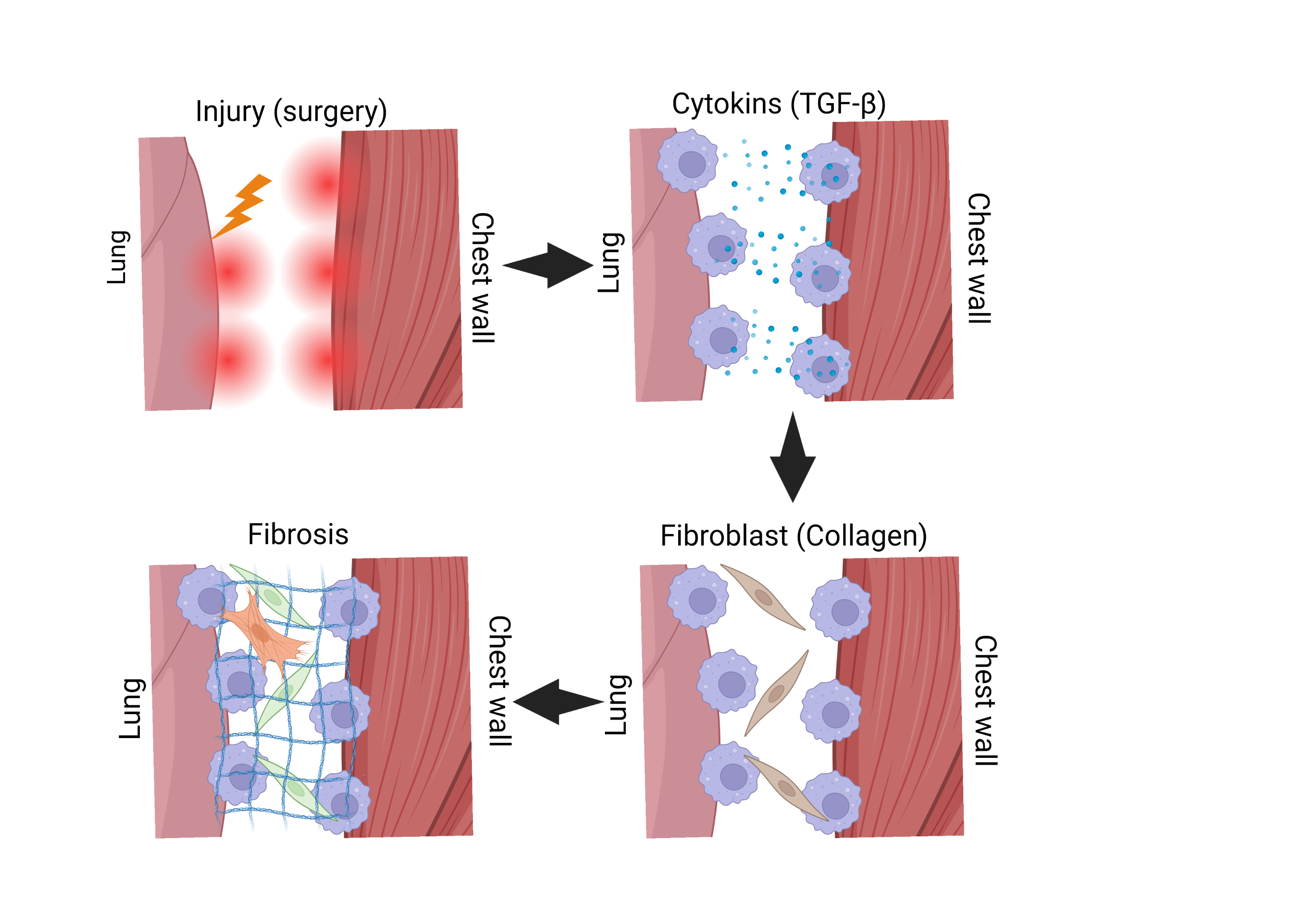

Intrathoracic adhesions can develop after thoracic surgery and increase the risk of complications during repeat surgery. However, the mechanisms underlying their formation remain poorly understood. In this study, we aimed to understand how these adhesions form and identify potential targets to prevent or reduce them. First, we established a mouse model of intrathoracic adhesions using talc. Through gene expression analysis, we found that macrophages, a type of immune cell, gather on the injured lung surface and contribute to the accumulation and activation of collagen-producing cells. Furthermore, when macrophages were removed using a drug, the accumulation of collagen-producing cells was reduced, and pleural adhesions were significantly decreased. These findings suggest that targeting macrophage-driven fibrosis may offer a new approach to preventing and treating intrathoracic adhesions.
【Mechanism of pleural adhesion formation】
Pleural adhesion formation following macrophage accumulation on the injured lung surface.
Yu Mori, Tomohisa Sakaue, Mikio Okazaki, Mie Kurata and Hironori Izutani
Intrathoracic adhesions can develop after thoracic surgery and increase the risk of complications during repeat surgery. However, the detailed molecular mechanisms underlying their formation have remained unclear. In this study, we aimed to establish an animal model that closely reproduces pleural adhesions seen in patients and to identify key cells and signaling pathways involved in adhesion formation. Our goal was to find new methods to prevent adhesions after thoracic surgery. First, we injected Unitalc, a medical-grade talc, into the chest cavity of mice under anesthesia and established suitable conditions to induce pleural adhesions between the lungs and chest wall. By day 9 after talc injection, the mice developed strong adhesions between the lung and surrounding tissues, connected by collagen-rich fibrous tissue. These adhesions were similar to those seen during repeat surgery in patients and were formed consistently in our mouse model.
Next, by combining tissue examination with gene expression analysis, we found that CD68-positive macrophages, a type of immune cell, gathered on the lung surface during the early stage of adhesion formation. This was followed by an increase in SERPINH1-positive collagen-producing cells. Both cell types were recruited to the affected area before strong adhesions developed. These findings suggested that macrophages may help attract and activate collagen-producing cells. To test this idea, we removed macrophages using clodronate liposomes. This treatment reduced the accumulation of SERPINH1-positive cells and significantly decreased pleural adhesion formation. These results showed that macrophages play a key role in the development of pleural adhesions.
Our findings provide a basis for developing new methods to prevent intrathoracic adhesions by targeting macrophages and may lead to future clinical applications. However, since this study was conducted in mice, further research is needed to determine whether these findings can be applied to humans. Schematic illustrations were created using BioRender (BioRender.com).
Advertisement