HS-AFM image of a 2:2 complex of HGF and full-length Met formed in living cell.
Cell-cell communication primarily occurs through the activation of transmembrane receptors such as growth factor and cytokine receptors. Understanding the activation mechanisms of growth factor receptors has been a hot topic in structural biology. Among these, Met, the receptor for hepatocyte growth factor (HGF), plays definitive roles in morphogenesis, tissue regeneration, and cancer metastasis. Met activation is initiated by HGF binding, which induces receptor dimerization. While high-resolution techniques such as X-ray crystallography and cryo-electron microscopy (cryo-EM) have provided valuable insights into this process, they have been limited to truncated forms of Met, leaving the structure of the physiological receptor dimer unresolved. As with other growth factor receptors, elucidating the structure of the full-length receptor dimer in its native cellular environment poses a significant challenge, due to the difficulty of isolating the receptor dimer from living cells and achieving high-resolution structural characterization.
In this study, the physiological Met dimer was chemically cross-linked in living cells prior to isolation. A multifaceted approach was employed to elucidate its structure and investigate the underlying dimerization mechanism. The full-length Met dimer was visualized at the single-protein level using high-speed atomic force microscopy (HS-AFM). To further characterize the HGF-Met complex, the researchers integrated split-luciferase complementary assays, cryo-EM of in vitro reconstituted HGF-Met ectodomain complexes, and molecular dynamics (MD) simulations.
HS-AFM imaging revealed that Met and HGF form a 2:2 complex, with two HGF molecules positioned at the periphery of the dimer. The SP domains of HGF interact with two closely associated Sema domains of Met, a configuration further supported by normal mode flexible fitting. IPT regions of Met are stably connected while exhibiting significant conformational flexibility. The split-luciferase-based detection of ectodomain dimerization and MD simulations demonstrated that this stable connection is mediated by dimerization of the membrane-proximal IPT4 domains of Met. While HGF binding to the Sema domain of Met is known to promote receptor association at the extracellular head region, this study uncovers a critical mechanism of Met activation: HGF not only facilitates head-to-head association but also drives the dimerization of the IPT4 domains, thereby stabilizing the Met dimer.
Beyond elucidating this key activation mechanism of Met receptor, this study also presents a powerful strategy for investigating physiological protein complexes. By combining in-cell cross-linking with HS-AFM, the dynamic structures of such complexes can be visualized at high spatiotemporal resolution. When integrated with computational analysis, this approach offers significant potential for advancing our understanding of complex biological systems, particularly those involving transient or heterogeneous multiprotein assemblies that are challenging to resolve using conventional methods.
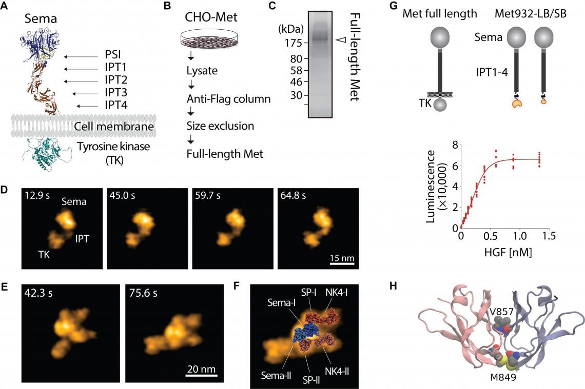
Figure 1. HGF-induced Met dimerization. The AlphaFold3-predicted structure of Met (A). Schematics of full-length Met purification (B). SDS-PAGE and silver staining of purified full-length Met (C). HS-AFM images of full-length Met on mica (D). HS-AFM images of 2:2 HGF-Met complex isolated from living cells (E). Normal mode flexible fitting of the atomistic model of the HGF-Sema structure to the HS-AFM image (F). Split-luciferase complementary assay demonstrating HGF-induced Met dimerization at IPT4 domain (G). Molecular model of an IPT4 dimer (H).
Contact
Kimie Nishimura (Ms.)
Project Planning and Outreach, NanoLSI Administration Office
Nano Life Science Institute, Kanazawa University
Kakuma-machi, Kanazawa 920-1192, Japan
Email: [email protected]
Nano Life Science Institute (WPI-NanoLSI), Kanazawa University
Understanding nanoscale mechanisms of life phenomena by exploring “uncharted nano-realms.” Cells are the basic units of life. At NanoLSI, researchers develop nanoprobe technologies that enable direct imaging, analysis, and manipulation of biomolecules such as proteins and nucleic acids inside living cells. By visualizing these processes at the nanoscale, the institute seeks to uncover fundamental principles of life and disease.
https://nanolsi.kanazawa-u.ac.jp/en/
About the World Premier International Research Center Initiative (WPI)
The WPI program was launched in 2007 by Japan’s Ministry of Education, Culture, Sports, Science and Technology (MEXT) to foster world-class research centers with outstanding research environments. WPI centers enjoy a high degree of autonomy, enabling innovative management and global collaboration. The program is administered by the Japan Society for the Promotion of Science (JSPS).
WPI News Portal: https://www.eurekalert.org/newsportal/WPI
Main WPI program site: www.jsps.go.jp/english/e-toplevel
About Kanazawa University
Founded in 1862 in Ishikawa Prefecture, Kanazawa University is one of Japan’s leading comprehensive national universities with a history spanning more than 160 years. With campuses at Kakuma and Takaramachi–Tsuruma, the university upholds its guiding principle of being “a research university dedicated to education, while opening its doors to both local and global society.”
Internationally recognized for its research institutes, including the Nano Life Science Institute (WPI-NanoLSI) and the Cancer Research Institute, Kanazawa University promotes interdisciplinary research and global collaboration, driving progress in health, sustainability, and culture.